


 癌症的基本特征包括细胞增殖、血管生成、迁移、凋亡逃避机制和细胞永生等。找到癌症发生过程中这些通路的关键标记物和对应的抗体用于检测至关重要。
 为您推荐一个泛素化位点预测神器——泛素化分析工具,可以为您的蛋白的泛素化位点作出预测和评分。
 细胞自噬受体图形绘图工具为你的蛋白的细胞受体结合位点作出预测和评分,识别结合到自噬通路中的蛋白是非常重要的,便于让我们理解自噬在正常生理、病理过程中的作用,如发育、细胞分化、神经退化性疾病、压力条件下、感染和癌症。
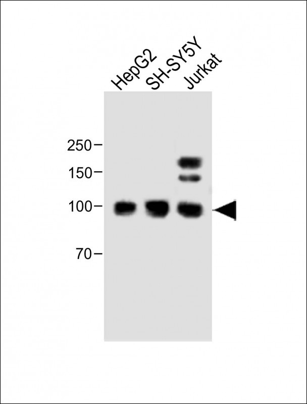
MGAT5 Antibody (C-term)
Affinity Purified Rabbit Polyclonal Antibody (Pab)
- 产品详情
- 实验流程
- 背景知识
Application
| WB, FC, E |
|---|---|
| Primary Accession | Q09328 |
| Other Accession | NP_002401.1 |
| Reactivity | Human |
| Host | Rabbit |
| Clonality | Polyclonal |
| Isotype | Rabbit IgG |
| Calculated MW | 84543 Da |
| Antigen Region | 652-680 aa |
| Gene ID | 4249 |
|---|---|
| Other Names | Alpha-1, 6-mannosylglycoprotein 6-beta-N-acetylglucosaminyltransferase A, Alpha-mannoside beta-1, 6-N-acetylglucosaminyltransferase, GlcNAc-T V, GNT-V, Mannoside acetylglucosaminyltransferase 5, N-acetylglucosaminyl-transferase V, MGAT5, GGNT5 |
| Target/Specificity | This MGAT5 antibody is generated from rabbits immunized with a KLH conjugated synthetic peptide between 652-680 amino acids from the C-terminal region of human MGAT5. |
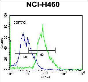
| Dilution | WB~~1:500 FC~~1:10~50 E~~Use at an assay dependent concentration. |
| Format | Purified polyclonal antibody supplied in PBS with 0.05% (V/V) Proclin 300. This antibody is purified through a protein A column, followed by peptide affinity purification. |
| Storage | Maintain refrigerated at 2-8°C for up to 2 weeks. For long term storage store at -20°C in small aliquots to prevent freeze-thaw cycles. |
| Precautions | MGAT5 Antibody (C-term) is for research use only and not for use in diagnostic or therapeutic procedures. |
| Name | MGAT5 |
|---|---|
| Synonyms | GGNT5 |
| Function | Catalyzes the addition of N-acetylglucosamine (GlcNAc) in beta 1-6 linkage to the alpha-linked mannose of biantennary N-linked oligosaccharides (PubMed:10395745, PubMed:30140003). Catalyzes an important step in the biosynthesis of branched, complex-type N-glycans, such as those found on EGFR, TGFR (TGF-beta receptor) and CDH2 (PubMed:10395745, PubMed:22614033, PubMed:30140003). Via its role in the biosynthesis of complex N-glycans, plays an important role in the activation of cellular signaling pathways, reorganization of the actin cytoskeleton, cell-cell adhesion and cell migration. MGAT5-dependent EGFR N-glycosylation enhances the interaction between EGFR and LGALS3 and thereby prevents rapid EGFR endocytosis and prolongs EGFR signaling. Required for efficient interaction between TGFB1 and its receptor. Enhances activation of intracellular signaling pathways by several types of growth factors, including FGF2, PDGF, IGF, TGFB1 and EGF. MGAT5-dependent CDH2 N-glycosylation inhibits CDH2-mediated homotypic cell-cell adhesion and contributes to the regulation of downstream signaling pathways. Promotes cell migration. Contributes to the regulation of the inflammatory response. MGAT5-dependent TCR N- glycosylation enhances the interaction between TCR and LGALS3, limits agonist-induced TCR clustering, and thereby dampens TCR-mediated responses to antigens. Required for normal leukocyte evasation and accumulation at sites of inflammation (By similarity). Inhibits attachment of monocytes to the vascular endothelium and subsequent monocyte diapedesis (PubMed:22614033). |
| Cellular Location | Golgi apparatus membrane {ECO:0000250|UniProtKB:P97259}; Single-pass type II membrane protein |
For Research Use Only. Not For Use In Diagnostic Procedures.
Provided below are standard protocols that you may find useful for product applications.
BACKGROUND
This gene encodes mannosyl (alpha-1,6-)-glycoprotein beta-1,6-N-acetyl-glucosaminyltransferase, a glycosyltransferase involved in the synthesis of protein-bound and lipid-bound oligosaccharides. Alterations of the oligosaccharides on cell surface glycoproteins cause significant changes in the adhesive or migratory behavior of a cell. Increase in the encoded protein's activity may correlate with the progression of invasive malignancies.
REFERENCES
Dick, D.M., et al. Am. J. Med. Genet. B Neuropsychiatr. Genet. 153B (6), 1179-1188 (2010) :
Brynedal, B., et al. J. Neuroimmunol. 220 (1-2), 120-124 (2010) :
Benson, V., et al. Int. Immunol. 22(3):167-177(2010)
Wang, C., et al. J. Cell. Biochem. 109(1):113-123(2010)
Ding, H., et al. Stroke 41(1):177-180(2010)
终于等到您。ABCEPTA(百远生物)抗体产品。
点击下方“我要评价 ”按钮提交您的反馈信息,您的反馈和评价是我们最宝贵的财富之一,
我们将在1-3个工作日内处理您的反馈信息。
如有疑问,联系:0512-88856768 tech-china@abcepta.com.


