


 癌症的基本特征包括细胞增殖、血管生成、迁移、凋亡逃避机制和细胞永生等。找到癌症发生过程中这些通路的关键标记物和对应的抗体用于检测至关重要。
 为您推荐一个泛素化位点预测神器——泛素化分析工具,可以为您的蛋白的泛素化位点作出预测和评分。
 细胞自噬受体图形绘图工具为你的蛋白的细胞受体结合位点作出预测和评分,识别结合到自噬通路中的蛋白是非常重要的,便于让我们理解自噬在正常生理、病理过程中的作用,如发育、细胞分化、神经退化性疾病、压力条件下、感染和癌症。
CDKN2B Antibody (C-term)
Affinity Purified Rabbit Polyclonal Antibody (Pab)
- 产品详情
- 实验流程
- 背景知识
Application
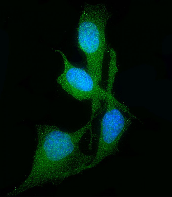
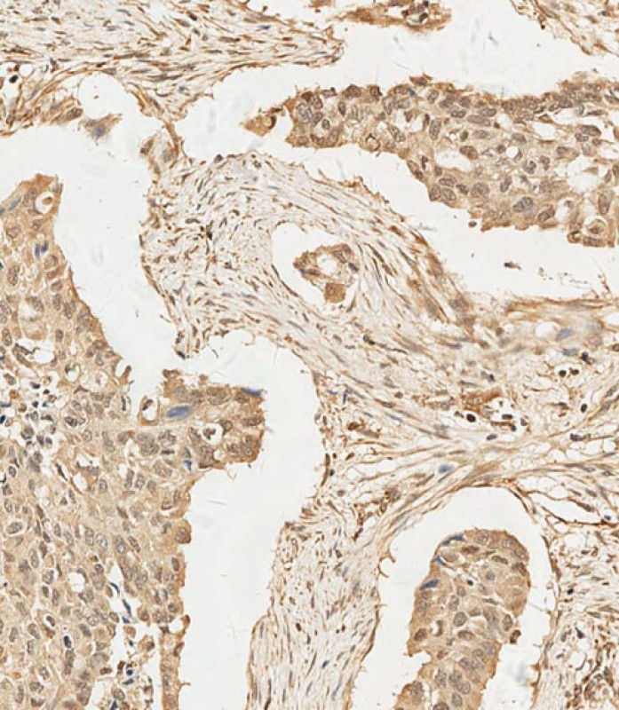
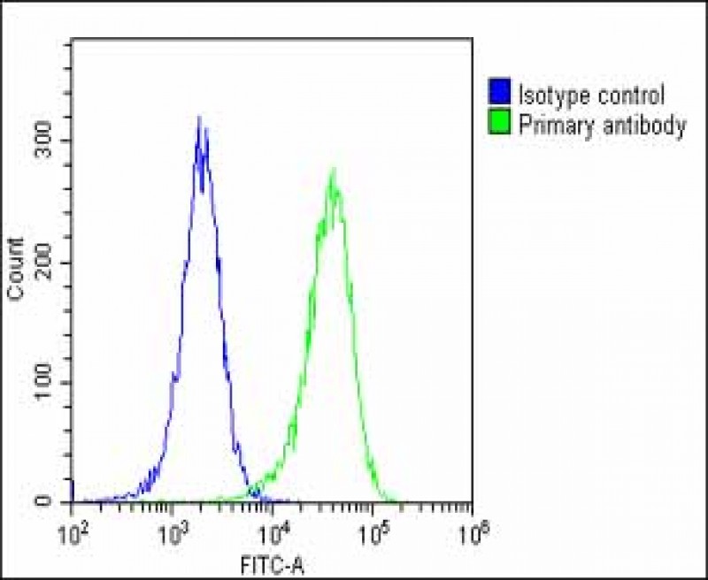
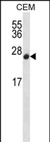
| IF, WB, FC, IHC-P, E |
|---|---|
| Primary Accession | P42772 |
| Other Accession | NP_004927.2, NP_511042.1 |
| Reactivity | Human |
| Host | Rabbit |
| Clonality | Polyclonal |
| Isotype | Rabbit IgG |
| Calculated MW | 14722 Da |
| Antigen Region | 103-131 aa |
| Gene ID | 1030 |
|---|---|
| Other Names | Cyclin-dependent kinase 4 inhibitor B, Multiple tumor suppressor 2, MTS-2, p14-INK4b, p15-INK4b, p15INK4B, CDKN2B, MTS2 |
| Target/Specificity | This CDKN2B antibody is generated from rabbits immunized with a KLH conjugated synthetic peptide between 103-131 amino acids from the C-terminal region of human CDKN2B. |
| Dilution | IF~~1:25 WB~~1:1000 FC~~1:25 IHC-P~~1:100 E~~Use at an assay dependent concentration. |
| Format | Purified polyclonal antibody supplied in PBS with 0.09% (W/V) sodium azide. This antibody is purified through a protein A column, followed by peptide affinity purification. |
| Storage | Maintain refrigerated at 2-8°C for up to 2 weeks. For long term storage store at -20°C in small aliquots to prevent freeze-thaw cycles. |
| Precautions | CDKN2B Antibody (C-term) is for research use only and not for use in diagnostic or therapeutic procedures. |
| Name | CDKN2B |
|---|---|
| Synonyms | MTS2 |
| Function | Interacts strongly with CDK4 and CDK6. Potent inhibitor. Potential effector of TGF-beta induced cell cycle arrest. |
| Cellular Location | Cytoplasm. Note=Also found in the nucleus |
| Tissue Location | Isoform 2 is expressed in normal (keratinocytes, fibroblasts) and tumor cell lines. |
For Research Use Only. Not For Use In Diagnostic Procedures.
Provided below are standard protocols that you may find useful for product applications.
BACKGROUND
This gene lies adjacent to the tumor suppressor gene CDKN2A in a region that is frequently mutated and deleted in a wide variety of tumors. This gene encodes a cyclin-dependent kinase inhibitor, which forms a complex with CDK4 or CDK6, and prevents the activation of the CDK kinases, thus the encoded protein functions as a cell growth regulator that controls cell cycle G1 progression. The expression of this gene was found to be dramatically induced by TGF beta, which suggested its role in the TGF beta induced growth inhibition. Two alternatively spliced transcript variants of this gene, which encode distinct proteins, have been reported.
REFERENCES
Camacho, C.V., et al. Carcinogenesis 31(10):1889-1896(2010)
Bailey, S.D., et al. Diabetes Care 33(10):2250-2253(2010)
Pechlivanis, S., et al. Arterioscler. Thromb. Vasc. Biol. 30(9):1867-1872(2010)
Heni, M., et al. Diabetes (2010) In press :
Roder, C., et al. Childs Nerv Syst (2010) In press :
终于等到您。ABCEPTA(百远生物)抗体产品。
点击下方“我要评价 ”按钮提交您的反馈信息,您的反馈和评价是我们最宝贵的财富之一,
我们将在1-3个工作日内处理您的反馈信息。
如有疑问,联系:0512-88856768 tech-china@abcepta.com.


