


 癌症的基本特征包括细胞增殖、血管生成、迁移、凋亡逃避机制和细胞永生等。找到癌症发生过程中这些通路的关键标记物和对应的抗体用于检测至关重要。
 为您推荐一个泛素化位点预测神器——泛素化分析工具,可以为您的蛋白的泛素化位点作出预测和评分。
 细胞自噬受体图形绘图工具为你的蛋白的细胞受体结合位点作出预测和评分,识别结合到自噬通路中的蛋白是非常重要的,便于让我们理解自噬在正常生理、病理过程中的作用,如发育、细胞分化、神经退化性疾病、压力条件下、感染和癌症。
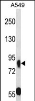
MUPCDH Antibody (N-term) 精选
Affinity Purified Rabbit Polyclonal Antibody (Pab)
- 产品详情
- 实验流程
- 背景知识
Application 
| WB, E |
|---|---|
| Primary Accession | Q9HBB8 |
| Other Accession | NP_001165439.1, NP_068743.2 |
| Reactivity | Human |
| Host | Rabbit |
| Clonality | Polyclonal |
| Isotype | Rabbit IgG |
| Calculated MW | 88223 Da |
| Antigen Region | 131-160 aa |
| Gene ID | 53841 |
|---|---|
| Other Names | Cadherin-related family member 5, Mu-protocadherin, Mucin and cadherin-like protein, CDHR5, MUCDHL, MUPCDH |
| Target/Specificity | This MUPCDH antibody is generated from rabbits immunized with a KLH conjugated synthetic peptide between 131-160 amino acids from the N-terminal region of human MUPCDH. |
| Dilution | WB~~1:1000 E~~Use at an assay dependent concentration. |
| Format | Purified polyclonal antibody supplied in PBS with 0.09% (W/V) sodium azide. This antibody is purified through a protein A column, followed by peptide affinity purification. |
| Storage | Maintain refrigerated at 2-8°C for up to 2 weeks. For long term storage store at -20°C in small aliquots to prevent freeze-thaw cycles. |
| Precautions | MUPCDH Antibody (N-term) is for research use only and not for use in diagnostic or therapeutic procedures. |
For Research Use Only. Not For Use In Diagnostic Procedures.
| Name | CDHR5 (HGNC:7521) |
|---|---|
| Function | Intermicrovillar adhesion molecule that forms, via its extracellular domain, calcium-dependent heterophilic complexes with CDHR2 on adjacent microvilli. Thereby, controls the packing of microvilli at the apical membrane of epithelial cells. Through its cytoplasmic domain, interacts with microvillus cytoplasmic proteins to form the intermicrovillar adhesion complex/IMAC. This complex plays a central role in microvilli and epithelial brush border differentiation. |
| Cellular Location | Apical cell membrane; Single-pass type I membrane protein. Cell projection, microvillus membrane; Single-pass type I membrane protein |
| Tissue Location | Highest expression in kidney, liver, colon and small intestine. In kidney, expressed apically along brush border of proximal convoluted tubule but not in cortical collecting ducts Isoform 1 is expressed primarily in adult small intestine and colon Isoform 2 is highly expressed in fetal liver (PubMed:12167596) Expressed in duodenum with higher expression in enterocytes along the villus axis and lower expression in crypts (at protein level) (PubMed:24725409). |
Provided below are standard protocols that you may find useful for product applications.
BACKGROUND
This gene is a novel mucin-like gene that is a member of the cadherin superfamily. While encoding nonpolymorphic tandem repeats rich in proline, serine and threonine similar to mucin proteins, the gene also contains sequence encoding calcium-binding motifs found in all cadherins. The role of the hybrid extracellular region and the specific function of this protein have not yet been determined. Alternatively spliced transcript variants encoding different isoforms have been described.
REFERENCES
Bailey, S.D., et al. Diabetes Care 33(10):2250-2253(2010)
Talmud, P.J., et al. Am. J. Hum. Genet. 85(5):628-642(2009)
Goldberg, M., et al. Hum. Genet. 112(4):334-342(2003)
Goldberg, M., et al. Am. J. Physiol. Renal Physiol. 283 (3), F454-F463 (2002) :
Paris, M.J., et al. Genomics 69(2):196-202(2000)

终于等到您。ABCEPTA(百远生物)抗体产品。
点击下方“我要评价 ”按钮提交您的反馈信息,您的反馈和评价是我们最宝贵的财富之一,
我们将在1-3个工作日内处理您的反馈信息。
如有疑问,联系:0512-88856768 tech-china@abcepta.com.


