


 癌症的基本特征包括细胞增殖、血管生成、迁移、凋亡逃避机制和细胞永生等。找到癌症发生过程中这些通路的关键标记物和对应的抗体用于检测至关重要。
 为您推荐一个泛素化位点预测神器——泛素化分析工具,可以为您的蛋白的泛素化位点作出预测和评分。
 细胞自噬受体图形绘图工具为你的蛋白的细胞受体结合位点作出预测和评分,识别结合到自噬通路中的蛋白是非常重要的,便于让我们理解自噬在正常生理、病理过程中的作用,如发育、细胞分化、神经退化性疾病、压力条件下、感染和癌症。
PPEF2 Antibody (Center)
Affinity Purified Rabbit Polyclonal Antibody (Pab)
- 产品详情
- 实验流程
- 背景知识
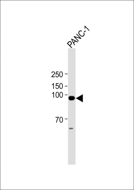
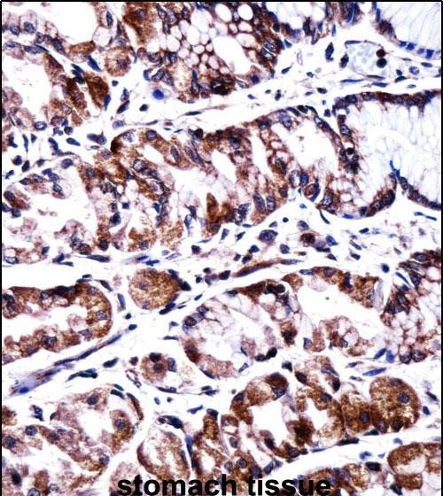
Application
| WB, IHC-P, E |
|---|---|
| Primary Accession | O14830 |
| Other Accession | NP_006230.2 |
| Reactivity | Human |
| Host | Rabbit |
| Clonality | Polyclonal |
| Isotype | Rabbit IgG |
| Calculated MW | 86518 Da |
| Antigen Region | 304-333 aa |
| Gene ID | 5470 |
|---|---|
| Other Names | Serine/threonine-protein phosphatase with EF-hands 2, PPEF-2, PPEF2 |
| Target/Specificity | This PPEF2 antibody is generated from rabbits immunized with a KLH conjugated synthetic peptide between 304-333 amino acids from the Central region of human PPEF2. |
| Dilution | WB~~1:2000 IHC-P~~1:100~500 E~~Use at an assay dependent concentration. |
| Format | Purified polyclonal antibody supplied in PBS with 0.05% (V/V) Proclin 300. This antibody is prepared by Saturated Ammonium Sulfate (SAS) precipitation followed by dialysis against PBS. |
| Storage | Maintain refrigerated at 2-8°C for up to 2 weeks. For long term storage store at -20°C in small aliquots to prevent freeze-thaw cycles. |
| Precautions | PPEF2 Antibody (Center) is for research use only and not for use in diagnostic or therapeutic procedures. |
| Name | PPEF2 |
|---|---|
| Function | May play a role in phototransduction. May dephosphorylate photoactivated rhodopsin. May function as a calcium sensing regulator of ionic currents, energy production or synaptic transmission. |
| Cellular Location | Cytoplasm. Cell projection, cilium, photoreceptor outer segment. Photoreceptor inner segment Note=Localized to photoreceptors, PPEF-2(L) is at least 2 fold more abundant in rod inner segments than in the outer segments |
| Tissue Location | Retinal specific. |
For Research Use Only. Not For Use In Diagnostic Procedures.
Provided below are standard protocols that you may find useful for product applications.
BACKGROUND
This gene encodes a member of the serine/threonine protein phosphatase with EF-hand motif family. The protein contains a protein phosphatase catalytic domain, and at least two EF-hand calcium-binding motifs in its C terminus. Although its substrate(s) is unknown, the encoded protein, which is expressed specifically in photoreceptors and the pineal, has been suggested to play a role in the visual system. This gene shares high sequence similarity with the Drosophila retinal degeneration C (rdgC) gene. [provided by RefSeq].
REFERENCES
Kutuzov, M.A., et al. Biochem. Biophys. Res. Commun. 293(3):1047-1052(2002)
Ramulu, P., et al. Mol. Cell. Biol. 21(24):8605-8614(2001)
Sherman, P.M., et al. Proc. Natl. Acad. Sci. U.S.A. 94(21):11639-11644(1997)
终于等到您。ABCEPTA(百远生物)抗体产品。
点击下方“我要评价 ”按钮提交您的反馈信息,您的反馈和评价是我们最宝贵的财富之一,
我们将在1-3个工作日内处理您的反馈信息。
如有疑问,联系:0512-88856768 tech-china@abcepta.com.


