


 癌症的基本特征包括细胞增殖、血管生成、迁移、凋亡逃避机制和细胞永生等。找到癌症发生过程中这些通路的关键标记物和对应的抗体用于检测至关重要。
 为您推荐一个泛素化位点预测神器——泛素化分析工具,可以为您的蛋白的泛素化位点作出预测和评分。
 细胞自噬受体图形绘图工具为你的蛋白的细胞受体结合位点作出预测和评分,识别结合到自噬通路中的蛋白是非常重要的,便于让我们理解自噬在正常生理、病理过程中的作用,如发育、细胞分化、神经退化性疾病、压力条件下、感染和癌症。
Mouse Plk3 Antibody (N-term)
Affinity Purified Rabbit Polyclonal Antibody (Pab)
- 产品详情
- 实验流程
- 背景知识
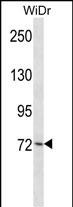
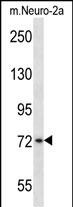
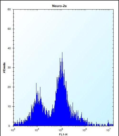
Application
| WB, FC, E |
|---|---|
| Primary Accession | Q60806 |
| Reactivity | Human, Rat, Mouse |
| Host | Rabbit |
| Clonality | Polyclonal |
| Isotype | Rabbit IgG |
| Calculated MW | 70012 Da |
| Antigen Region | 33-61 aa |
| Other Names | Serine/threonine-protein kinase PLK3, Cytokine-inducible serine/threonine-protein kinase, FGF-inducible kinase, Polo-like kinase 3, PLK-3, Plk3, Cnk, Fnk |
|---|---|
| Target/Specificity | This Mouse Plk3 antibody is generated from rabbits immunized with a KLH conjugated synthetic peptide between 33-61 amino acids from the N-terminal region of mouse Plk3. |
| Dilution | WB~~1:1000 FC~~1:10~50 E~~Use at an assay dependent concentration. |
| Format | Purified polyclonal antibody supplied in PBS with 0.09% (W/V) sodium azide. This antibody is purified through a protein A column, followed by peptide affinity purification. |
| Storage | Maintain refrigerated at 2-8°C for up to 2 weeks. For long term storage store at -20°C in small aliquots to prevent freeze-thaw cycles. |
| Precautions | Mouse Plk3 Antibody (N-term) is for research use only and not for use in diagnostic or therapeutic procedures. |
| Name | Plk3 |
|---|---|
| Synonyms | Cnk, Fnk |
| Function | Serine/threonine-protein kinase involved in cell cycle regulation, response to stress and Golgi disassembly. Polo-like kinases act by binding and phosphorylating proteins that are already phosphorylated on a specific motif recognized by the POLO box domains. Phosphorylates ATF2, BCL2L1, CDC25A, CDC25C, CHEK2, HIF1A, JUN, p53/TP53, p73/TP73, PTEN, TOP2A and VRK1. Involved in cell cycle regulation: required for entry into S phase and cytokinesis. Phosphorylates BCL2L1, leading to regulate the G2 checkpoint and progression to cytokinesis during mitosis. Plays a key role in response to stress: rapidly activated upon stress stimulation, such as ionizing radiation, reactive oxygen species (ROS), hyperosmotic stress, UV irradiation and hypoxia. Involved in DNA damage response and G1/S transition checkpoint by phosphorylating CDC25A, p53/TP53 and p73/TP73. Phosphorylates p53/TP53 in response to reactive oxygen species (ROS), thereby promoting p53/TP53-mediated apoptosis. Phosphorylates CHEK2 in response to DNA damage, promoting the G2/M transition checkpoint. Phosphorylates the transcription factor p73/TP73 in response to DNA damage, leading to inhibit p73/TP73-mediated transcriptional activation and pro-apoptotic functions. Phosphorylates HIF1A and JUN is response to hypoxia. Phosphorylates ATF2 following hyperosmotic stress in corneal epithelium. Also involved in Golgi disassembly during the cell cycle: part of a MEK1/MAP2K1-dependent pathway that induces Golgi fragmentation during mitosis by mediating phosphorylation of VRK1. May participate in endomitotic cell cycle, a form of mitosis in which both karyokinesis and cytokinesis are interrupted and is a hallmark of megakaryocyte differentiation, via its interaction with CIB1. |
| Cellular Location | Cytoplasm. Nucleus. Nucleus, nucleolus. Golgi apparatus Cytoplasm, cytoskeleton, microtubule organizing center, centrosome. Note=Translocates to the nucleus upon cisplatin treatment. Localizes to the Golgi apparatus during interphase (By similarity). |
| Tissue Location | Expressed in skin. |
Research Areas
For Research Use Only. Not For Use In Diagnostic Procedures.
Application Protocols
Provided below are standard protocols that you may find useful for product applications.
BACKGROUND
Serine/threonine protein kinase involved in regulating M phase functions during the cell cycle. May also be part of the signaling network controlling cellular adhesion. In vitro, is able to phosphorylate CDC25C and casein (By similarity).
终于等到您。ABCEPTA(百远生物)抗体产品。
点击下方“我要评价 ”按钮提交您的反馈信息,您的反馈和评价是我们最宝贵的财富之一,
我们将在1-3个工作日内处理您的反馈信息。
如有疑问,联系:0512-88856768 tech-china@abcepta.com.
¥ 1,250.00
Cat# AP14695a


