


 癌症的基本特征包括细胞增殖、血管生成、迁移、凋亡逃避机制和细胞永生等。找到癌症发生过程中这些通路的关键标记物和对应的抗体用于检测至关重要。
 为您推荐一个泛素化位点预测神器——泛素化分析工具,可以为您的蛋白的泛素化位点作出预测和评分。
 细胞自噬受体图形绘图工具为你的蛋白的细胞受体结合位点作出预测和评分,识别结合到自噬通路中的蛋白是非常重要的,便于让我们理解自噬在正常生理、病理过程中的作用,如发育、细胞分化、神经退化性疾病、压力条件下、感染和癌症。
Alkaline Phosphatase (ALPI) Antibody (Center) 精选
Purified Rabbit Polyclonal Antibody (Pab)

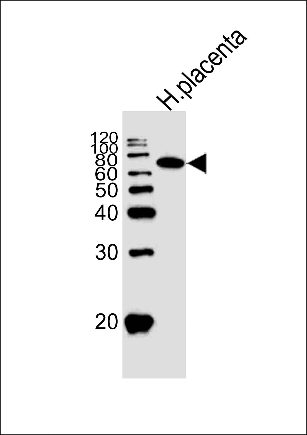
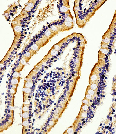
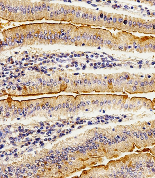
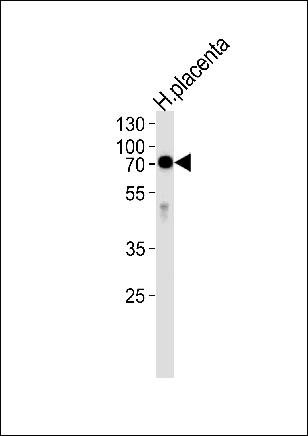
_(Center)_-_HeLa.jpg)
- 产品详情
- 文献引用 : 1
- 实验流程
- 背景知识
Application 
| IF, WB, IHC-P, E |
|---|---|
| Primary Accession | P09923 |
| Reactivity | Human |
| Host | Rabbit |
| Clonality | Polyclonal |
| Isotype | Rabbit IgG |
| Calculated MW | 56812 Da |
| Antigen Region | 274-304 aa |
| Gene ID | 248 |
|---|---|
| Other Names | Intestinal-type alkaline phosphatase, IAP, Intestinal alkaline phosphatase, ALPI |
| Target/Specificity | This Alkaline Phosphatase (ALPI) antibody is generated from rabbits immunized with a KLH conjugated synthetic peptide between 274-304 amino acids from the Central region of human Alkaline Phosphatase (ALPI). |
| Dilution | IF~~1:100 WB~~1:1000 IHC-P~~1:100 E~~Use at an assay dependent concentration. |
| Format | Purified polyclonal antibody supplied in PBS with 0.09% (W/V) sodium azide. This antibody is prepared by Saturated Ammonium Sulfate (SAS) precipitation followed by dialysis against PBS. |
| Storage | Maintain refrigerated at 2-8°C for up to 2 weeks. For long term storage store at -20°C in small aliquots to prevent freeze-thaw cycles. |
| Precautions | Alkaline Phosphatase (ALPI) Antibody (Center) is for research use only and not for use in diagnostic or therapeutic procedures. |
For Research Use Only. Not For Use In Diagnostic Procedures.
| Name | ALPI |
|---|---|
| Function | Alkaline phosphatase that can hydrolyze various phosphate compounds. |
| Cellular Location | Cell membrane {ECO:0000250|UniProtKB:P15693}; Lipid-anchor, GPI-anchor {ECO:0000250|UniProtKB:P15693} |

Provided below are standard protocols that you may find useful for product applications.
BACKGROUND
There are at least four distinct but related alkaline phosphatases: intestinal, placental, placental-like, and liver/bone/kidney (tissue non-specific). The intestinal alkaline phosphatase gene encodes a digestive brush-border enzyme. This enzyme is upregulated during small intestinal epithelial cell differentiation.
REFERENCES
References for protein:
1.Torres,M.I., Pathol. Res. Pract. 203 (6), 485-487 (2007)
2.Olsen,L., Am. J. Physiol. Gastrointest. Liver Physiol. 289 (2), G220-G226(2005)
3.Alkhoury,F., Am. J. Physiol. Gastrointest. Liver Physiol. 289 (2), G285-G290(2005)
References for HeLa cell line:
1. Scherer WF, Syverton JT, Gey GO (May 1953). "Studies on the propagation in vitro of poliomyelitis viruses. IV. Viral multiplication in a stable strain of human malignant epithelial cells (strain HeLa) derived from an epidermoid carcinoma of the cervix". J. Exp. Med. 97 (5): 695–710. [PubMed:13052828].
2. Macville M, Schröck E, Padilla-Nash H, Keck C, Ghadimi BM, Zimonjic D, Popescu N, Ried T (January 1999). "Comprehensive and definitive molecular cytogenetic characterization of HeLa cells by spectral karyotyping". Cancer Res. 59 (1): 141–50. [PubMed: 9892199].
3. Rahbari R, Sheahan T, Modes V, Collier P, Macfarlane C, Badge RM (April 2009). "A novel L1 retrotransposon marker for HeLa cell line identification". BioTechniques 46 (4): 277–84. [PubMed: 19450234].
4. Capes-Davis A, Theodosopoulos G, Atkin I, Drexler HG, Kohara A, MacLeod RA, Masters JR, Nakamura Y, Reid YA, Reddel RR, Freshney RI (July 2010). "Check your cultures! A list of cross-contaminated or misidentified cell lines". Int. J. Cancer 127 (1): 1–8. [PubMed:20143388].

终于等到您。ABCEPTA(百远生物)抗体产品。
点击下方“我要评价 ”按钮提交您的反馈信息,您的反馈和评价是我们最宝贵的财富之一,
我们将在1-3个工作日内处理您的反馈信息。
如有疑问,联系:0512-88856768 tech-china@abcepta.com.


