


 癌症的基本特征包括细胞增殖、血管生成、迁移、凋亡逃避机制和细胞永生等。找到癌症发生过程中这些通路的关键标记物和对应的抗体用于检测至关重要。
 为您推荐一个泛素化位点预测神器——泛素化分析工具,可以为您的蛋白的泛素化位点作出预测和评分。
 细胞自噬受体图形绘图工具为你的蛋白的细胞受体结合位点作出预测和评分,识别结合到自噬通路中的蛋白是非常重要的,便于让我们理解自噬在正常生理、病理过程中的作用,如发育、细胞分化、神经退化性疾病、压力条件下、感染和癌症。
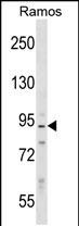
PPP1R9B Antibody (N-term) 精选
Affinity Purified Rabbit Polyclonal Antibody (Pab)
- 产品详情
- 实验流程
- 背景知识
Application 
| WB, E |
|---|---|
| Primary Accession | Q96SB3 |
| Other Accession | NP_115984.3 |
| Reactivity | Human |
| Predicted | Mouse, Rat, Bovine, Canine |
| Host | Rabbit |
| Clonality | Polyclonal |
| Isotype | Rabbit IgG |
| Calculated MW | 89334 Da |
| Antigen Region | 24-53 aa |
| Gene ID | 84687 |
|---|---|
| Other Names | Neurabin-2, Neurabin-II, Protein phosphatase 1 regulatory subunit 9B, Spinophilin, PPP1R9B, PPP1R6 |
| Target/Specificity | This PPP1R9B antibody is generated from rabbits immunized with a KLH conjugated synthetic peptide between 24-53 amino acids from the N-terminal region of human PPP1R9B. |
| Dilution | WB~~1:1000 E~~Use at an assay dependent concentration. |
| Format | Purified polyclonal antibody supplied in PBS with 0.09% (W/V) sodium azide. This antibody is purified through a protein A column, followed by peptide affinity purification. |
| Storage | Maintain refrigerated at 2-8°C for up to 2 weeks. For long term storage store at -20°C in small aliquots to prevent freeze-thaw cycles. |
| Precautions | PPP1R9B Antibody (N-term) is for research use only and not for use in diagnostic or therapeutic procedures. |
For Research Use Only. Not For Use In Diagnostic Procedures.
| Name | PPP1R9B |
|---|---|
| Synonyms | PPP1R6 |
| Function | Seems to act as a scaffold protein in multiple signaling pathways. Modulates excitatory synaptic transmission and dendritic spine morphology. Binds to actin filaments (F-actin) and shows cross- linking activity. Binds along the sides of the F-actin. May play an important role in linking the actin cytoskeleton to the plasma membrane at the synaptic junction. Believed to target protein phosphatase 1/PP1 to dendritic spines, which are rich in F-actin, and regulates its specificity toward ion channels and other substrates, such as AMPA-type and NMDA-type glutamate receptors. Plays a role in regulation of G protein-coupled receptor signaling, including dopamine D2 receptors and alpha-adrenergic receptors. May establish a signaling complex for dopaminergic neurotransmission through D2 receptors by linking receptors downstream signaling molecules and the actin cytoskeleton. Binds to ADRA1B and RGS2 and mediates regulation of ADRA1B signaling. May confer to Rac signaling specificity by binding to both, RacGEFs and Rac effector proteins. Probably regulates p70 S6 kinase activity by forming a complex with TIAM1 (By similarity). Required for hepatocyte growth factor (HGF)-induced cell migration. |
| Cellular Location | Cytoplasm, cytoskeleton. Nucleus. Cell projection, dendritic spine {ECO:0000250|UniProtKB:O35274}. Postsynaptic density {ECO:0000250|UniProtKB:O35274}. Synapse. Cell junction, adherens junction. Cytoplasm. Cell membrane. Cell projection, lamellipodium. Cell projection, filopodium. Cell projection, ruffle membrane. Note=Enriched at synapse and cadherin-based cell-cell adhesion sites. In neurons, both cytosolic and membrane-associated, and highly enriched in the postsynaptic density apposed to exitatory synapses. Colocalizes with PPP1R2 at actin-rich adherens junctions in epithelial cells and in dendritic spines (By similarity). Accumulates in the lamellipodium, filopodium and ruffle membrane in response to hepatocyte growth factor (HGF) treatment. |
Provided below are standard protocols that you may find useful for product applications.
BACKGROUND
Spinophilin is a regulatory subunit of protein phosphatase-1 catalytic subunit (PP1; see MIM 176875) and is highly enriched in dendritic spines, specialized protrusions from dendritic shafts that receive most of the excitatory input in the central nervous system (Allen et al., 1997 [PubMed 9275233]).
REFERENCES
Pinheiro, A.P., et al. Am. J. Med. Genet. B Neuropsychiatr. Genet. 153B (5), 1070-1080 (2010) :
Rajagopal, S., et al. J. Biol. Chem. 285(23):18060-18071(2010)
Martins-de-Souza, D., et al. Eur Arch Psychiatry Clin Neurosci 259(3):151-163(2009)
Sagara, M., et al. Oncogene 28(10):1357-1365(2009)
Meng, X., et al. Eur. J. Immunol. 39(2):552-560(2009)

终于等到您。ABCEPTA(百远生物)抗体产品。
点击下方“我要评价 ”按钮提交您的反馈信息,您的反馈和评价是我们最宝贵的财富之一,
我们将在1-3个工作日内处理您的反馈信息。
如有疑问,联系:0512-88856768 tech-china@abcepta.com.


