


 癌症的基本特征包括细胞增殖、血管生成、迁移、凋亡逃避机制和细胞永生等。找到癌症发生过程中这些通路的关键标记物和对应的抗体用于检测至关重要。
 为您推荐一个泛素化位点预测神器——泛素化分析工具,可以为您的蛋白的泛素化位点作出预测和评分。
 细胞自噬受体图形绘图工具为你的蛋白的细胞受体结合位点作出预测和评分,识别结合到自噬通路中的蛋白是非常重要的,便于让我们理解自噬在正常生理、病理过程中的作用,如发育、细胞分化、神经退化性疾病、压力条件下、感染和癌症。
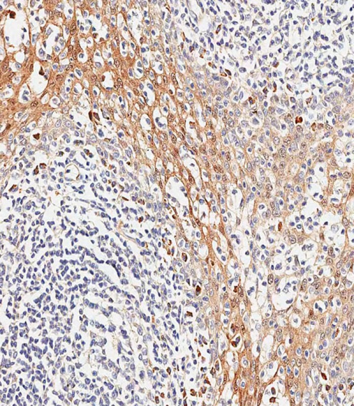
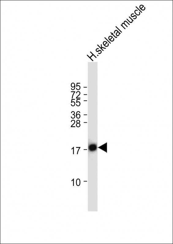
TSC22D3 Antibody (Center)
Affinity Purified Rabbit Polyclonal Antibody (Pab)
- 产品详情
- 实验流程
- 背景知识
Application
| IHC-P-Leica, WB, E |
|---|---|
| Primary Accession | Q99576 |
| Other Accession | Q9EQZ1, P80220, NP_004080.2, NP_001015881.1 |
| Reactivity | Human, Rat, Mouse |
| Predicted | Rat, Pig |
| Host | Rabbit |
| Clonality | Polyclonal |
| Isotype | Rabbit IgG |
| Calculated MW | 14810 Da |
| Antigen Region | 62-91 aa |
| Gene ID | 1831 |
|---|---|
| Other Names | TSC22 domain family protein 3, DSIP-immunoreactive peptide, Protein DIP, hDIP, Delta sleep-inducing peptide immunoreactor, Glucocorticoid-induced leucine zipper protein, GILZ, TSC-22-like protein, TSC-22-related protein, TSC-22R, TSC22D3, DSIPI, GILZ |
| Target/Specificity | This TSC22D3 antibody is generated from rabbits immunized with a KLH conjugated synthetic peptide between 62-91 amino acids from the Central region of human TSC22D3. |
| Dilution | IHC-P-Leica~~1:500 WB~~1:2000 E~~Use at an assay dependent concentration. |
| Format | Purified polyclonal antibody supplied in PBS with 0.09% (W/V) sodium azide. This antibody is purified through a protein A column, followed by peptide affinity purification. |
| Storage | Maintain refrigerated at 2-8°C for up to 2 weeks. For long term storage store at -20°C in small aliquots to prevent freeze-thaw cycles. |
| Precautions | TSC22D3 Antibody (Center) is for research use only and not for use in diagnostic or therapeutic procedures. |
| Name | TSC22D3 (HGNC:3051) |
|---|---|
| Function | Protects T-cells from IL2 deprivation-induced apoptosis through the inhibition of FOXO3A transcriptional activity that leads to the down-regulation of the pro-apoptotic factor BCL2L11 (PubMed:15031210). In macrophages, plays a role in the anti- inflammatory and immunosuppressive effects of glucocorticoids and IL10 (PubMed:12393603). In T-cells, inhibits anti-CD3-induced NFKB1 nuclear translocation and thereby NFKB1 DNA-binding activities (PubMed:11468175). In vitro, suppresses AP-1 transcription factor complex DNA-binding activities (By similarity). |
| Cellular Location | [Isoform 1]: Cytoplasm {ECO:0000250|UniProtKB:Q9Z2S7}. Nucleus {ECO:0000250|UniProtKB:Q9Z2S7} Note=Localization depends on differentiation status of myoblasts (By similarity). In undifferentiated myoblasts; localizes to the cytoplasm, but in differentiating myoblast; localizes to the nucleus (By similarity). {ECO:0000250|UniProtKB:Q9Z2S7} |
| Tissue Location | Ubiquitously expressed, including in the fetal brain and liver (PubMed:26752201). Expressed in brain, lung, spleen and skeletal muscle (PubMed:11313722, PubMed:12393603). Lower levels detected in heart and kidney (PubMed:11313722, PubMed:12393603). Not detected in the pancreas (PubMed:11313722). In non-lymphoid tissues, in the absence of inflammation, the major source of constitutive expression is the macrophage lineage (PubMed:12393603). Also expressed in cells from different hemopoietic cell lineages, including bone marrow cells, CD34+ stem cells, mature B- and T-cells, monocytes and granulocytes (PubMed:11313722). Down-regulated in activated macrophages from inflammatory lesions of delayed-type hypersensitivity (DTH) reactions, such as in tuberculosis and in Crohn disease, whereas in Burkitt lymphoma, persists in macrophages involved in the phagocytosis of apoptotic malignant cells (PubMed:12393603) |
For Research Use Only. Not For Use In Diagnostic Procedures.
Provided below are standard protocols that you may find useful for product applications.
BACKGROUND
The protein encoded by this gene shares significant sequence identity with the murine TSC-22 and Drosophila shs, both of which are leucine zipper proteins, that function as transcriptional regulators. The expression of this gene is stimulated by glucocorticoids and interleukin 10, and it appears to play a key role in the anti-inflammatory and immunosuppressive effects of this steroid and chemokine. Transcript variants encoding different isoforms have been identified for this gene. [provided by RefSeq].
REFERENCES
Latre de Late, P., et al. J. Biol. Chem. 285(8):5594-5605(2010)
Lekva, T., et al. J. Clin. Endocrinol. Metab. 95(1):246-255(2010)
Soundararajan, R., et al. Proc. Natl. Acad. Sci. U.S.A. 106(19):7804-7809(2009)
Zhang, X.H., et al. Clin. Exp. Allergy 39(5):647-654(2009)
Redjimi, N., et al. Mol. Cancer 8, 83 (2009) :
终于等到您。ABCEPTA(百远生物)抗体产品。
点击下方“我要评价 ”按钮提交您的反馈信息,您的反馈和评价是我们最宝贵的财富之一,
我们将在1-3个工作日内处理您的反馈信息。
如有疑问,联系:0512-88856768 tech-china@abcepta.com.


