


 癌症的基本特征包括细胞增殖、血管生成、迁移、凋亡逃避机制和细胞永生等。找到癌症发生过程中这些通路的关键标记物和对应的抗体用于检测至关重要。
 为您推荐一个泛素化位点预测神器——泛素化分析工具,可以为您的蛋白的泛素化位点作出预测和评分。
 细胞自噬受体图形绘图工具为你的蛋白的细胞受体结合位点作出预测和评分,识别结合到自噬通路中的蛋白是非常重要的,便于让我们理解自噬在正常生理、病理过程中的作用,如发育、细胞分化、神经退化性疾病、压力条件下、感染和癌症。
TESK1 Antibody (Center)
Affinity Purified Rabbit Polyclonal Antibody (Pab)
- 产品详情
- 实验流程
- 背景知识
Application
| WB, E |
|---|---|
| Primary Accession | Q15569 |
| Other Accession | Q63572, NP_006276.2 |
| Reactivity | Human, Rat, Mouse |
| Predicted | Rat |
| Host | Rabbit |
| Clonality | Polyclonal |
| Isotype | Rabbit IgG |
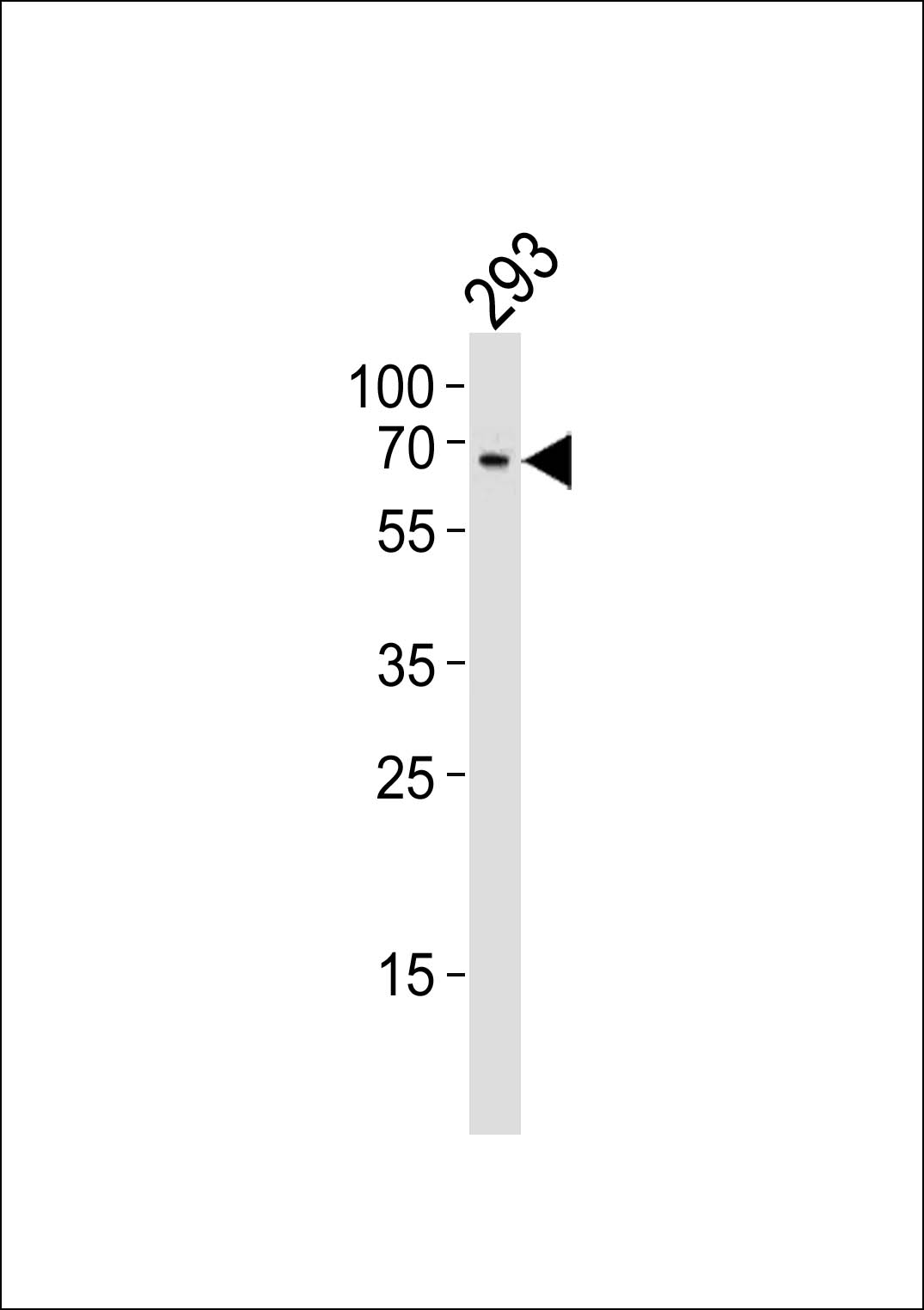
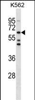
| Calculated MW | 67684 Da |
| Antigen Region | 187-215 aa |
| Gene ID | 7016 |
|---|---|
| Other Names | Dual specificity testis-specific protein kinase 1, Testicular protein kinase 1, TESK1 |
| Target/Specificity | This TESK1 antibody is generated from rabbits immunized with a KLH conjugated synthetic peptide between 187-215 amino acids from the Central region of human TESK1. |
| Dilution | WB~~1:1000 E~~Use at an assay dependent concentration. |
| Format | Purified polyclonal antibody supplied in PBS with 0.09% (W/V) sodium azide. This antibody is purified through a protein A column, followed by peptide affinity purification. |
| Storage | Maintain refrigerated at 2-8°C for up to 2 weeks. For long term storage store at -20°C in small aliquots to prevent freeze-thaw cycles. |
| Precautions | TESK1 Antibody (Center) is for research use only and not for use in diagnostic or therapeutic procedures. |
| Name | TESK1 |
|---|---|
| Function | Dual specificity protein kinase activity catalyzing autophosphorylation and phosphorylation of exogenous substrates on both serine/threonine and tyrosine residues (By similarity). Regulates the cellular cytoskeleton by enhancing actin stress fiber formation via phosphorylation of cofilin and by preventing microtubule breakdown via inhibition of TAOK1/MARKK kinase activity (By similarity). Inhibits podocyte motility via regulation of actin cytoskeletal dynamics and phosphorylation of CFL1 (By similarity). Positively regulates integrin- mediated cell spreading, via phosphorylation of cofilin (PubMed:15584898). Suppresses ciliogenesis via multiple pathways; phosphorylation of CFL1, suppression of ciliary vesicle directional trafficking to the ciliary base, and by facilitating YAP1 nuclear localization where it acts as a transcriptional corepressor of the TEAD4 target genes AURKA and PLK1 (PubMed:25849865). Probably plays a central role at and after the meiotic phase of spermatogenesis (By similarity). |
| Cellular Location | Cytoplasm. Cytoplasm, perinuclear region {ECO:0000250|UniProtKB:Q63572} Cytoplasm, cytoskeleton, microtubule organizing center, centrosome. Cell projection, lamellipodium {ECO:0000250|UniProtKB:Q63572}. Note=Colocalizes with SPRY4 in vesicular spots in the cytoplasm (PubMed:15584898). Localized to F- actin-rich lamellipodia at the cell periphery following fibronectin- mediated cell adhesion of Schwann cells (By similarity) {ECO:0000250|UniProtKB:Q63572, ECO:0000269|PubMed:15584898} |
| Tissue Location | Expressed in podocytes and renal tubular cells in the kidney (at protein level). |
For Research Use Only. Not For Use In Diagnostic Procedures.
Provided below are standard protocols that you may find useful for product applications.
BACKGROUND
This gene product is a serine/threonine protein kinase that contains an N-terminal protein kinase domain and a C-terminal proline-rich domain. Its protein kinase domain is most closely related to those of the LIM motif-containing protein kinases (LIMKs). The encoded protein can phosphorylate myelin basic protein and histone in vitro. The testicular germ cell-specific expression and developmental pattern of expression of the mouse gene suggests that this gene plays an important role at and after the meiotic phase of spermatogenesis.
REFERENCES
Davila, S., et al. Genes Immun. 11(3):232-238(2010)
Johne, C., et al. Mol. Biol. Cell 19(4):1391-1403(2008)
LaLonde, D.P., et al. J. Biol. Chem. 280(22):21680-21688(2005)
Leeksma, O.C., et al. Eur. J. Biochem. 269(10):2546-2556(2002)
Toshima, J.Y., et al. J. Biol. Chem. 276(46):43471-43481(2001)
终于等到您。ABCEPTA(百远生物)抗体产品。
点击下方“我要评价 ”按钮提交您的反馈信息,您的反馈和评价是我们最宝贵的财富之一,
我们将在1-3个工作日内处理您的反馈信息。
如有疑问,联系:0512-88856768 tech-china@abcepta.com.


