


 癌症的基本特征包括细胞增殖、血管生成、迁移、凋亡逃避机制和细胞永生等。找到癌症发生过程中这些通路的关键标记物和对应的抗体用于检测至关重要。
 为您推荐一个泛素化位点预测神器——泛素化分析工具,可以为您的蛋白的泛素化位点作出预测和评分。
 细胞自噬受体图形绘图工具为你的蛋白的细胞受体结合位点作出预测和评分,识别结合到自噬通路中的蛋白是非常重要的,便于让我们理解自噬在正常生理、病理过程中的作用,如发育、细胞分化、神经退化性疾病、压力条件下、感染和癌症。
RAB28 Antibody (Center)
Affinity Purified Rabbit Polyclonal Antibody (Pab)
- 产品详情
- 实验流程
- 背景知识
Application
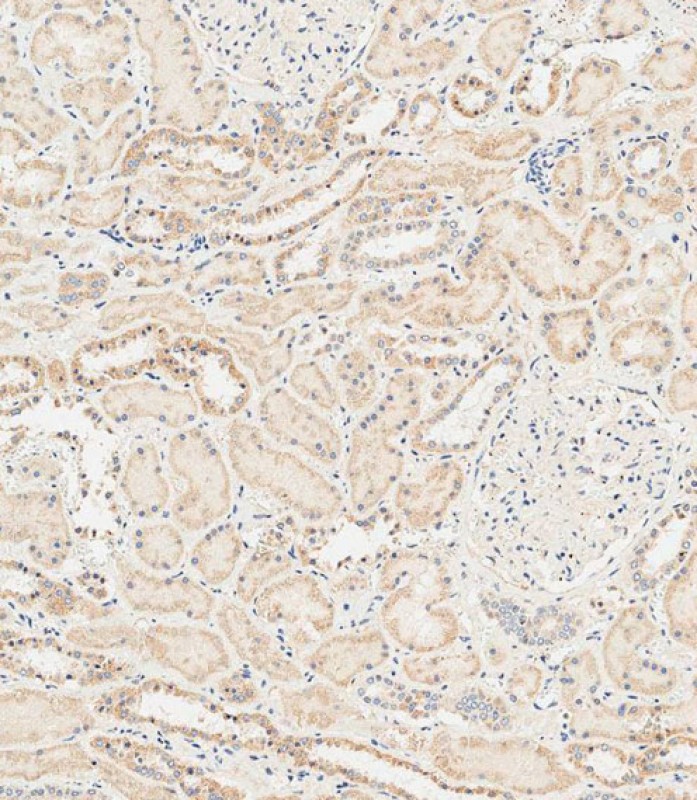
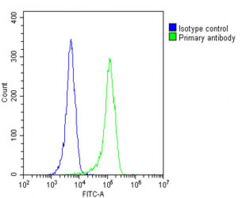
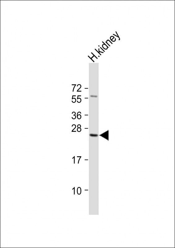
| WB, FC, IHC-P-Leica, E |
|---|---|
| Primary Accession | P51157 |
| Other Accession | NP_004240.2, NP_001017979.1 |
| Reactivity | Human, Rat, Mouse |
| Host | Rabbit |
| Clonality | Polyclonal |
| Isotype | Rabbit IgG |
| Calculated MW | 24841 Da |
| Antigen Region | 119-147 aa |
| Gene ID | 9364 |
|---|---|
| Other Names | Ras-related protein Rab-28, RAB28 |
| Target/Specificity | This RAB28 antibody is generated from rabbits immunized with a KLH conjugated synthetic peptide between 119-147 amino acids from the Central region of human RAB28. |
| Dilution | WB~~1:1000 FC~~1:25 IHC-P-Leica~~1:500 E~~Use at an assay dependent concentration. |
| Format | Purified polyclonal antibody supplied in PBS with 0.09% (W/V) sodium azide. This antibody is purified through a protein A column, followed by peptide affinity purification. |
| Storage | Maintain refrigerated at 2-8°C for up to 2 weeks. For long term storage store at -20°C in small aliquots to prevent freeze-thaw cycles. |
| Precautions | RAB28 Antibody (Center) is for research use only and not for use in diagnostic or therapeutic procedures. |
| Name | RAB28 (HGNC:9768) |
|---|---|
| Function | The small GTPases Rab are key regulators of intracellular membrane trafficking, from the formation of transport vesicles to their fusion with membranes (PubMed:8647132). Rabs cycle between an inactive GDP-bound form and an active GTP-bound form that is able to recruit to membranes different sets of downstream effectors directly responsible for vesicle formation, movement, tethering and fusion (PubMed:8647132). RAB28 is required for shedding and phagocytosis of cone cell outer segments (OS) discs in the retina (By similarity). Also participates in nuclear factor kappa-B p65/RELA nuclear transport in endothelial cells (By similarity). |
| Cellular Location | Cell membrane; Lipid-anchor; Cytoplasmic side. Cytoplasm, cytoskeleton, cilium basal body {ECO:0000250|UniProtKB:P51158}. Cytoplasm {ECO:0000250|UniProtKB:P51158}. Nucleus {ECO:0000250|UniProtKB:P51158} Note=Expressed in the basal body and ciliary rootlet of the photoreceptor cells (By similarity). Localized in the cytoplasm and the nucleus of vascular endothelial cells (By similarity) {ECO:0000250|UniProtKB:P51158} |
| Tissue Location | [Isoform S]: Isoform S is detected in most tissues investigated: cortex, liver, kidney, skeletal muscle, adipose tissue, testis, urothelium, lung, bone marrow and retinal pigment epithelium (RPE). [Isoform 3]: Isoform 3 is highly expressed in heart, lung, bone marrow, retina, brain, and RPE |
For Research Use Only. Not For Use In Diagnostic Procedures.
Provided below are standard protocols that you may find useful for product applications.
BACKGROUND
This gene encodes a member of the Rab subfamily of Ras-related small GTPases. The encoded protein may be involved in regulating intracellular trafficking. Alternative splicing results in multiple transcript variants. Pseudogenes of this gene are found on chromosomes 9 and X.
REFERENCES
Rose, J.E., et al. Mol. Med. 16 (7-8), 247-253 (2010) :
Lee, S.H., et al. FEBS Lett. 582(29):4107-4111(2008)
Lamesch, P., et al. Genomics 89(3):307-315(2007)
Stenmark, H., et al. Genome Biol. 2 (5), REVIEWS3007 (2001) :
Pereira-Leal, J.B., et al. J. Mol. Biol. 301(4):1077-1087(2000)
终于等到您。ABCEPTA(百远生物)抗体产品。
点击下方“我要评价 ”按钮提交您的反馈信息,您的反馈和评价是我们最宝贵的财富之一,
我们将在1-3个工作日内处理您的反馈信息。
如有疑问,联系:0512-88856768 tech-china@abcepta.com.


