


 癌症的基本特征包括细胞增殖、血管生成、迁移、凋亡逃避机制和细胞永生等。找到癌症发生过程中这些通路的关键标记物和对应的抗体用于检测至关重要。
 为您推荐一个泛素化位点预测神器——泛素化分析工具,可以为您的蛋白的泛素化位点作出预测和评分。
 细胞自噬受体图形绘图工具为你的蛋白的细胞受体结合位点作出预测和评分,识别结合到自噬通路中的蛋白是非常重要的,便于让我们理解自噬在正常生理、病理过程中的作用,如发育、细胞分化、神经退化性疾病、压力条件下、感染和癌症。
CD1D Antibody (Center)
Affinity Purified Rabbit Polyclonal Antibody (Pab)
- 产品详情
- 实验流程
- 背景知识
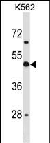
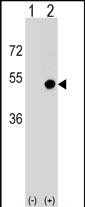
Application
| WB, E |
|---|---|
| Primary Accession | P15813 |
| Other Accession | NP_001757.1 |
| Reactivity | Human |
| Host | Rabbit |
| Clonality | Polyclonal |
| Isotype | Rabbit IgG |
| Calculated MW | 37717 Da |
| Antigen Region | 177-206 aa |
| Gene ID | 912 |
|---|---|
| Other Names | Antigen-presenting glycoprotein CD1d, R3G1, CD1d, CD1D |
| Target/Specificity | This CD1D antibody is generated from rabbits immunized with a KLH conjugated synthetic peptide between 177-206 amino acids from the Central region of human CD1D. |
| Dilution | WB~~1:1000 E~~Use at an assay dependent concentration. |
| Format | Purified polyclonal antibody supplied in PBS with 0.09% (W/V) sodium azide. This antibody is purified through a protein A column, followed by peptide affinity purification. |
| Storage | Maintain refrigerated at 2-8°C for up to 2 weeks. For long term storage store at -20°C in small aliquots to prevent freeze-thaw cycles. |
| Precautions | CD1D Antibody (Center) is for research use only and not for use in diagnostic or therapeutic procedures. |
| Name | CD1D |
|---|---|
| Function | Antigen-presenting protein that binds self and non-self glycolipids and presents them to T-cell receptors on natural killer T- cells. |
| Cellular Location | Cell membrane; Single-pass type I membrane protein. Basolateral cell membrane; Single-pass type I membrane protein. Endosome membrane; Single-pass type I membrane protein. Lysosome membrane; Single-pass type I membrane protein. Endoplasmic reticulum membrane; Single-pass type I membrane protein. Note=Subject to intracellular trafficking between the cell membrane, endosomes and lysosomes. |
| Tissue Location | Expressed on cortical thymocytes, on certain T-cell leukemias, and in various other tissues |
For Research Use Only. Not For Use In Diagnostic Procedures.
Provided below are standard protocols that you may find useful for product applications.
BACKGROUND
This gene encodes a divergent member of the CD1 family of transmembrane glycoproteins, which are structurally related to the major histocompatibility complex (MHC) proteins and form heterodimers with beta-2-microglobulin. The CD1 proteins mediate the presentation of primarily lipid and glycolipid antigens of self or microbial origin to T cells. The human genome contains five CD1 family genes organized in a cluster on chromosome 1. The CD1 family members are thought to differ in their cellular localization and specificity for particular lipid ligands. The protein encoded by this gene localizes to late endosomes and lysosomes via a tyrosine-based motif in the cytoplasmic tail.
REFERENCES
Miura, S., et al. J. Virol. 84(22):11614-11623(2010)
Moll, M., et al. Blood 116(11):1876-1884(2010)
Liu, J., et al. J. Immunol. 184(9):4973-4981(2010)
Davila, S., et al. Genes Immun. 11(3):232-238(2010)
Brandl, C., et al. PLoS ONE 5 (5), E10800 (2010) :
终于等到您。ABCEPTA(百远生物)抗体产品。
点击下方“我要评价 ”按钮提交您的反馈信息,您的反馈和评价是我们最宝贵的财富之一,
我们将在1-3个工作日内处理您的反馈信息。
如有疑问,联系:0512-88856768 tech-china@abcepta.com.


