


 癌症的基本特征包括细胞增殖、血管生成、迁移、凋亡逃避机制和细胞永生等。找到癌症发生过程中这些通路的关键标记物和对应的抗体用于检测至关重要。
 为您推荐一个泛素化位点预测神器——泛素化分析工具,可以为您的蛋白的泛素化位点作出预测和评分。
 细胞自噬受体图形绘图工具为你的蛋白的细胞受体结合位点作出预测和评分,识别结合到自噬通路中的蛋白是非常重要的,便于让我们理解自噬在正常生理、病理过程中的作用,如发育、细胞分化、神经退化性疾病、压力条件下、感染和癌症。
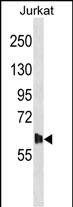
DMPK Antibody (N-term) 精选
Affinity Purified Rabbit Polyclonal Antibody (Pab)
- 产品详情
- 实验流程
- 背景知识
Application 
| WB, E |
|---|---|
| Primary Accession | Q09013 |
| Other Accession | NP_001075029.1, NP_001075031.1 |
| Reactivity | Human |
| Host | Rabbit |
| Clonality | Polyclonal |
| Isotype | Rabbit IgG |
| Calculated MW | 69385 Da |
| Antigen Region | 11-39 aa |
| Gene ID | 1760 |
|---|---|
| Other Names | Myotonin-protein kinase, MT-PK, DM-kinase, DMK, DM1 protein kinase, DMPK, Myotonic dystrophy protein kinase, DMPK, DM1PK, MDPK |
| Target/Specificity | This DMPK antibody is generated from rabbits immunized with a KLH conjugated synthetic peptide between 11-39 amino acids from the N-terminal region of human DMPK. |
| Dilution | WB~~1:1000 E~~Use at an assay dependent concentration. |
| Format | Purified polyclonal antibody supplied in PBS with 0.09% (W/V) sodium azide. This antibody is purified through a protein A column, followed by peptide affinity purification. |
| Storage | Maintain refrigerated at 2-8°C for up to 2 weeks. For long term storage store at -20°C in small aliquots to prevent freeze-thaw cycles. |
| Precautions | DMPK Antibody (N-term) is for research use only and not for use in diagnostic or therapeutic procedures. |
For Research Use Only. Not For Use In Diagnostic Procedures.
| Name | DMPK |
|---|---|
| Synonyms | DM1PK, MDPK |
| Function | Non-receptor serine/threonine protein kinase which is necessary for the maintenance of skeletal muscle structure and function. May play a role in myocyte differentiation and survival by regulating the integrity of the nuclear envelope and the expression of muscle-specific genes. May also phosphorylate PPP1R12A and inhibit the myosin phosphatase activity to regulate myosin phosphorylation. Also critical to the modulation of cardiac contractility and to the maintenance of proper cardiac conduction activity probably through the regulation of cellular calcium homeostasis. Phosphorylates PLN, a regulator of calcium pumps and may regulate sarcoplasmic reticulum calcium uptake in myocytes. May also phosphorylate FXYD1/PLM which is able to induce chloride currents. May also play a role in synaptic plasticity. |
| Cellular Location | Endoplasmic reticulum membrane; Single-pass type IV membrane protein; Cytoplasmic side. Nucleus outer membrane; Single-pass type IV membrane protein; Cytoplasmic side Mitochondrion outer membrane; Single-pass type IV membrane protein. Sarcoplasmic reticulum membrane. Cell membrane. Cytoplasm, cytosol. Note=Localizes to sarcoplasmic reticulum membranes of cardiomyocytes. [Isoform 3]: Mitochondrion membrane. |
| Tissue Location | Most isoforms are expressed in many tissues including heart, skeletal muscle, liver and brain, except for isoform 2 which is only found in the heart and skeletal muscle, and isoform 14 which is only found in the brain, with high levels in the striatum, cerebellar cortex and pons. |
Provided below are standard protocols that you may find useful for product applications.
BACKGROUND
The protein encoded by this gene is a serine-threonine kinase that is closely related to other kinases that interact with members of the Rho family of small GTPases. Substrates for this enzyme include myogenin, the beta-subunit of the L-type calcium channels, and phospholemman. The 3' untranslated region of this gene contains 5-37 copies of a CTG trinucleotide repeat. Expansion of this unstable motif to 50-5,000 copies causes myotonic dystrophy type I, which increases in severity with increasing repeat element copy number. Repeat expansion is associated with condensation of local chromatin structure that disrupts the expression of genes in this region. Several alternatively spliced transcript variants of this gene have been described, but the full-length nature of some of these variants has not been determined.
REFERENCES
Theerasasawat, S., et al. J Clin Neurosci 17(12):1520-1522(2010)
Santoro, M., et al. Exp. Mol. Pathol. 89(2):158-168(2010)
Bailey, S.D., et al. Diabetes Care 33(10):2250-2253(2010)
Vignaud, A., et al. Neuromuscul. Disord. 20(5):319-325(2010)
Kwon, M.J., et al. Ann. Clin. Lab. Sci. 40(2):156-162(2010)

终于等到您。ABCEPTA(百远生物)抗体产品。
点击下方“我要评价 ”按钮提交您的反馈信息,您的反馈和评价是我们最宝贵的财富之一,
我们将在1-3个工作日内处理您的反馈信息。
如有疑问,联系:0512-88856768 tech-china@abcepta.com.


