


 癌症的基本特征包括细胞增殖、血管生成、迁移、凋亡逃避机制和细胞永生等。找到癌症发生过程中这些通路的关键标记物和对应的抗体用于检测至关重要。
 为您推荐一个泛素化位点预测神器——泛素化分析工具,可以为您的蛋白的泛素化位点作出预测和评分。
 细胞自噬受体图形绘图工具为你的蛋白的细胞受体结合位点作出预测和评分,识别结合到自噬通路中的蛋白是非常重要的,便于让我们理解自噬在正常生理、病理过程中的作用,如发育、细胞分化、神经退化性疾病、压力条件下、感染和癌症。
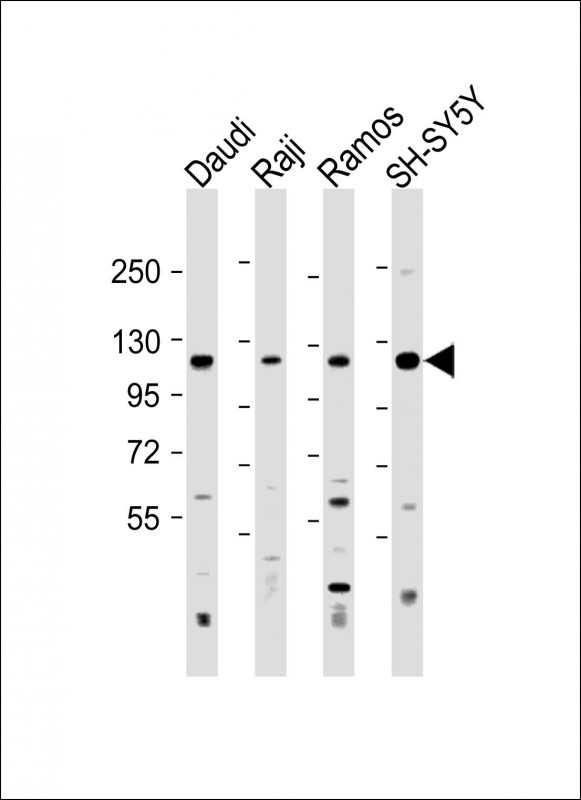
LILRB1 Antibody (Center)
Affinity Purified Rabbit Polyclonal Antibody (Pab)
- 产品详情
- 实验流程
- 背景知识
Application
| WB, E |
|---|---|
| Primary Accession | Q8NHL6 |
| Other Accession | NP_001075106.1, NP_001075107.1 |
| Reactivity | Human |
| Host | Rabbit |
| Clonality | Polyclonal |
| Isotype | Rabbit IgG |
| Calculated MW | 70819 Da |
| Antigen Region | 345-374 aa |
| Gene ID | 10859 |
|---|---|
| Other Names | Leukocyte immunoglobulin-like receptor subfamily B member 1, LIR-1, Leukocyte immunoglobulin-like receptor 1, CD85 antigen-like family member J, Immunoglobulin-like transcript 2, ILT-2, Monocyte/macrophage immunoglobulin-like receptor 7, MIR-7, CD85j, LILRB1, ILT2, LIR1, MIR7 |
| Target/Specificity | This LILRB1 antibody is generated from rabbits immunized with a KLH conjugated synthetic peptide between 345-374 amino acids from the Central region of human LILRB1. |
| Dilution | WB~~1:500-1:2000 E~~Use at an assay dependent concentration. |
| Format | Purified polyclonal antibody supplied in PBS with 0.09% (W/V) sodium azide. This antibody is purified through a protein A column, followed by peptide affinity purification. |
| Storage | Maintain refrigerated at 2-8°C for up to 2 weeks. For long term storage store at -20°C in small aliquots to prevent freeze-thaw cycles. |
| Precautions | LILRB1 Antibody (Center) is for research use only and not for use in diagnostic or therapeutic procedures. |
| Name | LILRB1 {ECO:0000303|PubMed:20600445, ECO:0000312|HGNC:HGNC:6605} |
|---|---|
| Function | Receptor for class I MHC antigens. Recognizes a broad spectrum of HLA-A, HLA-B, HLA-C, HLA-G and HLA-F alleles (PubMed:16455647, PubMed:28636952). Receptor for H301/UL18, a human cytomegalovirus class I MHC homolog. Ligand binding results in inhibitory signals and down-regulation of the immune response. Engagement of LILRB1 present on natural killer cells or T-cells by class I MHC molecules protects the target cells from lysis. Interaction with HLA-B or HLA-E leads to inhibition of FCER1A signaling and serotonin release. Inhibits FCGR1A-mediated phosphorylation of cellular proteins and mobilization of intracellular calcium ions (PubMed:11907092, PubMed:9285411, PubMed:9842885). Recognizes HLA-G in complex with B2M/beta-2 microglobulin and a nonamer self-peptide (PubMed:16455647). Upon interaction with peptide-bound HLA-G-B2M complex, triggers secretion of growth-promoting factors by decidual NK cells (PubMed:19304799, PubMed:29262349). Reprograms B cells toward an immune suppressive phenotype (PubMed:24453251). |
| Cellular Location | Cell membrane; Single-pass type I membrane protein |
| Tissue Location | Expressed in B cells, monocytes and various dendritic cell (DC) subsets including myeloid, plasmacytoid and tolerogenic DCs (at protein level) (PubMed:20448110, PubMed:24453251, PubMed:9285411, PubMed:9842885). Expressed in decidual macrophages (at protein level) (PubMed:19304799). Expressed in decidual NK cells (at protein level) (PubMed:29262349). |
For Research Use Only. Not For Use In Diagnostic Procedures.
Provided below are standard protocols that you may find useful for product applications.
BACKGROUND
This gene is a member of the leukocyte immunoglobulin-like receptor (LIR) family, which is found in a gene cluster at chromosomal region 19q13.4. The encoded protein belongs to the subfamily B class of LIR receptors which contain two or four extracellular immunoglobulin domains, a transmembrane domain, and two to four cytoplasmic immunoreceptor tyrosine-based inhibitory motifs (ITIMs). The receptor is expressed on immune cells where it binds to MHC class I molecules on antigen-presenting cells and transduces a negative signal that inhibits stimulation of an immune response. It is thought to control inflammatory responses and cytotoxicity to help focus the immune response and limit autoreactivity. Multiple transcript variants encoding different isoforms have been found for this gene.
REFERENCES
Bailey, S.D., et al. Diabetes Care 33(10):2250-2253(2010)
Davidson, C.L., et al. Hum. Immunol. 71(10):942-949(2010)
Huang, J., et al. J. Virol. 84(18):9463-9471(2010)
Godal, R., et al. Biol. Blood Marrow Transplant. 16(5):612-621(2010)
Lamar, D.L., et al. Blood 115(16):3278-3286(2010)
终于等到您。ABCEPTA(百远生物)抗体产品。
点击下方“我要评价 ”按钮提交您的反馈信息,您的反馈和评价是我们最宝贵的财富之一,
我们将在1-3个工作日内处理您的反馈信息。
如有疑问,联系:0512-88856768 tech-china@abcepta.com.


