


 癌症的基本特征包括细胞增殖、血管生成、迁移、凋亡逃避机制和细胞永生等。找到癌症发生过程中这些通路的关键标记物和对应的抗体用于检测至关重要。
 为您推荐一个泛素化位点预测神器——泛素化分析工具,可以为您的蛋白的泛素化位点作出预测和评分。
 细胞自噬受体图形绘图工具为你的蛋白的细胞受体结合位点作出预测和评分,识别结合到自噬通路中的蛋白是非常重要的,便于让我们理解自噬在正常生理、病理过程中的作用,如发育、细胞分化、神经退化性疾病、压力条件下、感染和癌症。
MR1 Antibody (C-term)
Affinity Purified Rabbit Polyclonal Antibody (Pab)
- 产品详情
- 实验流程
- 背景知识
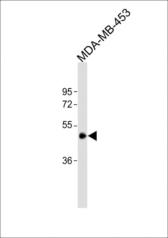
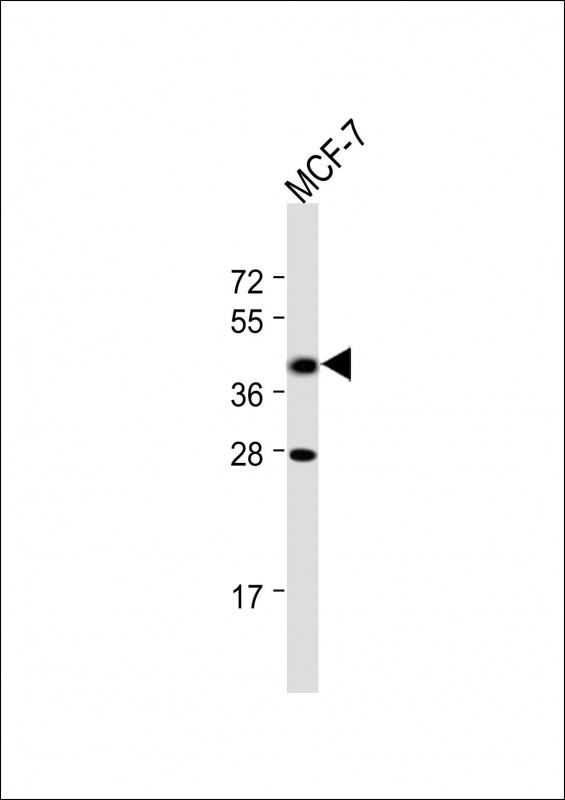
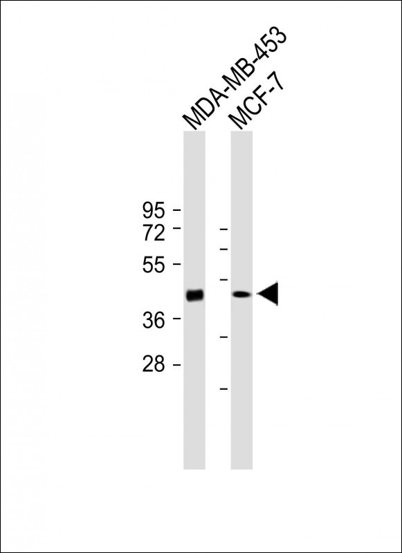
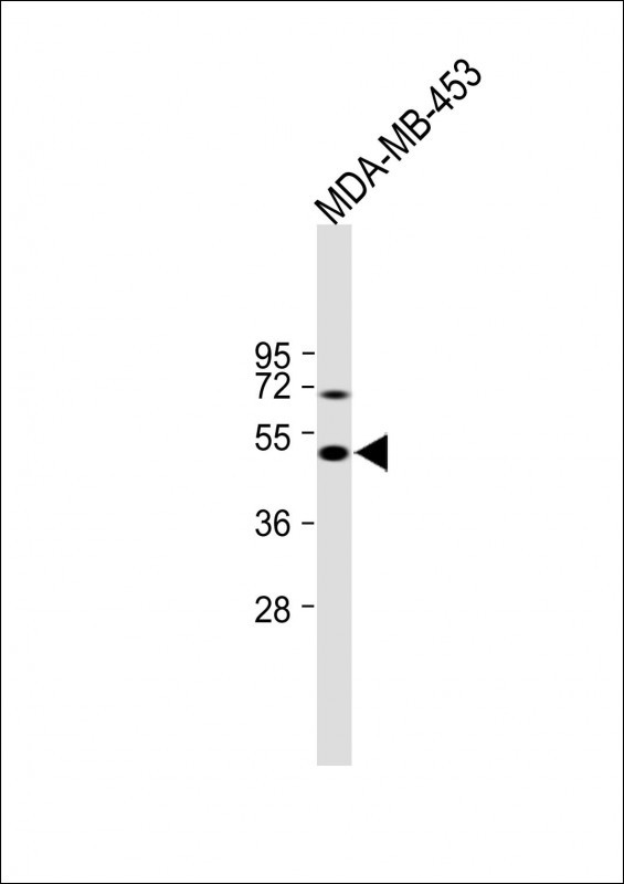
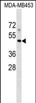
Application
| WB, E |
|---|---|
| Primary Accession | Q95460 |
| Other Accession | NP_001181929.1, NP_001181928.1 |
| Reactivity | Human |
| Host | Rabbit |
| Clonality | Polyclonal |
| Isotype | Rabbit IgG |
| Calculated MW | 39366 Da |
| Antigen Region | 312-341 aa |
| Gene ID | 3140 |
|---|---|
| Other Names | Major histocompatibility complex class I-related gene protein, MHC class I-related gene protein, Class I histocompatibility antigen-like protein, MR1 |
| Target/Specificity | This MR1 antibody is generated from rabbits immunized with a KLH conjugated synthetic peptide between 312-341 amino acids from the C-terminal region of human MR1. |
| Dilution | WB~~1:1000 E~~Use at an assay dependent concentration. |
| Format | Purified polyclonal antibody supplied in PBS with 0.05% (V/V) Proclin 300. This antibody is purified through a protein A column, followed by peptide affinity purification. |
| Storage | Maintain refrigerated at 2-8°C for up to 2 weeks. For long term storage store at -20°C in small aliquots to prevent freeze-thaw cycles. |
| Precautions | MR1 Antibody (C-term) is for research use only and not for use in diagnostic or therapeutic procedures. |
| Name | MR1 {ECO:0000303|PubMed:19416870, ECO:0000312|HGNC:HGNC:4975} |
|---|---|
| Function | Antigen-presenting molecule specialized in displaying microbial pyrimidine-based metabolites to alpha-beta T cell receptors (TCR) on innate-type mucosal-associated invariant T (MAIT) cells (PubMed:19416870, PubMed:23457030, PubMed:22692454, PubMed:23051753, PubMed:24101382, PubMed:23846752, PubMed:26795251). In complex with B2M preferentially presents riboflavin-derived metabolites to semi- invariant TRAV1.2 TCRs on MAIT cells, guiding immune surveillance of the microbial metabolome at mucosal epithelial barriers (PubMed:20581831, PubMed:24101382, PubMed:24695216, PubMed:26795251). Signature pyrimidine-based microbial antigens are generated via non- enzymatic condensation of metabolite intermediates of the riboflavin pathway with by-products arising from other metabolic pathways such as glycolysis. Typical potent antigenic metabolites are 5-(2- oxoethylideneamino)-6-D-ribitylaminouracil (5-OE-RU) and 5-(2- oxopropylideneamino)-6-D-ribitylaminouracil (5-OP-RU), products of condensation of 5-amino-6-D-ribityaminouracil (5-A-RU) with glyoxal or methylglyoxal by-products, respectively (PubMed:24695216, PubMed:32958637, PubMed:32709702). May present microbial antigens to various TRAV1-2-negative MAIT cell subsets, providing for unique recognition of diverse microbes, including pathogens that do not synthesize riboflavin (PubMed:27527800, PubMed:31113973). Upon antigen recognition, elicits rapid innate-type MAIT cell activation to eliminate pathogenic microbes by directly killing infected cells (PubMed:23846752, PubMed:24695216, PubMed:27527800). During T cell development, drives thymic selection and post-thymic terminal differentiation of MAIT cells in a process dependent on commensal microflora (By similarity). Acts as an immune sensor of cancer cell metabolome (PubMed:31959982). May present a tumor-specific or -associated metabolite essential for cancer cell survival to a 'pan- cancer' TCR consisting of TRAV38.2-DV8*TRAJ31 alpha chain paired with a TRBV25.1*TRBJ2.3 beta chain on a non-MAIT CD8-positive T cell clone (MC.7.G5), triggering T cell-mediated killing of a wide range of cancer cell types (PubMed:31959982). |
| Cellular Location | Cell membrane; Single-pass type I membrane protein Endoplasmic reticulum membrane; Single-pass type I membrane protein. Golgi apparatus membrane; Single-pass type I membrane protein. Early endosome membrane; Single-pass type I membrane protein. Late endosome membrane; Single-pass type I membrane protein. Note=In the absence of antigen remains within the endoplasmic reticulum where it acts as a metabolite sensor. Antigen binding triggers trafficking of the ternary complex to the plasma membrane. After presentation, most of these complexes are rapidly internalized and degraded via endocytosis. A small subset recycles via endosomes back to the plasma membrane and may thus acquire and present new antigens that do not efficiently reach the endoplasmic reticulum. [Isoform 3]: Cell membrane; Single-pass type I membrane protein. Endoplasmic reticulum membrane; Single-pass membrane protein. Note=The larger proportion remains in the ER in an immature state. The subset that reach cell surface does it through a B2M-independent pathway. |
| Tissue Location | Ubiquitous (PubMed:7624800, PubMed:9780177). Low expression is detected in peripheral blood B cells, T cells, monocytes and in bronchial epithelial cells (at protein level) (PubMed:27043408) Expressed in plasmablasts or plasma B cells in the lamina propria of ileum, appendix and colon (at protein level) (PubMed:19760593). Highly expressed on a subset of CD45-positive CD3-positive thymocytes (at protein level) (PubMed:22692454). |
For Research Use Only. Not For Use In Diagnostic Procedures.
Provided below are standard protocols that you may find useful for product applications.
BACKGROUND
MR1 has antigen presentation function. Involved in the development and expansion of a small population of T cells expressing an invariant T cell receptor alpha chain called mucosal-associated invariant T cells (MAIT). MAIT cells are preferentially located in the gut lamina propria and therfore may be involed in monitoring commensal flora or serve as a distress signal. Expression and MAIT cell recognition seem to be ligand-dependent.
REFERENCES
Gozalbo-Lopez, B., et al. Histol. Histopathol. 24(11):1439-1449(2009)
Stumpf, A.N., et al. Blood 114(17):3684-3692(2009)
Huang, S., et al. Proc. Natl. Acad. Sci. U.S.A. 106(20):8290-8295(2009)
Aldemir, H. Biochem. Biophys. Res. Commun. 366(2):328-334(2008)
Miley, M.J., et al. J. Immunol. 170(12):6090-6098(2003)
终于等到您。ABCEPTA(百远生物)抗体产品。
点击下方“我要评价 ”按钮提交您的反馈信息,您的反馈和评价是我们最宝贵的财富之一,
我们将在1-3个工作日内处理您的反馈信息。
如有疑问,联系:0512-88856768 tech-china@abcepta.com.


