


 癌症的基本特征包括细胞增殖、血管生成、迁移、凋亡逃避机制和细胞永生等。找到癌症发生过程中这些通路的关键标记物和对应的抗体用于检测至关重要。
 为您推荐一个泛素化位点预测神器——泛素化分析工具,可以为您的蛋白的泛素化位点作出预测和评分。
 细胞自噬受体图形绘图工具为你的蛋白的细胞受体结合位点作出预测和评分,识别结合到自噬通路中的蛋白是非常重要的,便于让我们理解自噬在正常生理、病理过程中的作用,如发育、细胞分化、神经退化性疾病、压力条件下、感染和癌症。
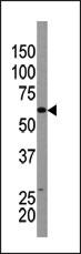
ATG9A Antibody (N-term)
Purified Rabbit Polyclonal Antibody (Pab)

- 产品详情
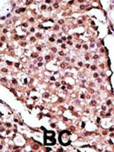
- 文献引用 : 1
- 实验流程
- 背景知识
Application
| WB, IHC-P, E |
|---|---|
| Primary Accession | Q7Z3C6 |
| Other Accession | Q5FWU3, Q68FE2, Q3T904 |
| Reactivity | Human, Mouse |
| Predicted | Bovine, Rat |
| Host | Rabbit |
| Clonality | Polyclonal |
| Isotype | Rabbit IgG |
| Calculated MW | 94447 Da |
| Antigen Region | 1-30 aa |
| Gene ID | 79065 |
|---|---|
| Other Names | Autophagy-related protein 9A, APG9-like 1, mATG9, ATG9A, APG9L1 |
| Target/Specificity | This ATG9A antibody is generated from rabbits immunized with a KLH conjugated synthetic peptide between 1-30 amino acids from the N-terminal region of human ATG9A. |
| Dilution | WB~~1:1000 IHC-P~~1:100~500 E~~Use at an assay dependent concentration. |
| Format | Purified polyclonal antibody supplied in PBS with 0.09% (W/V) sodium azide. This antibody is prepared by Saturated Ammonium Sulfate (SAS) precipitation followed by dialysis against PBS. |
| Storage | Maintain refrigerated at 2-8°C for up to 2 weeks. For long term storage store at -20°C in small aliquots to prevent freeze-thaw cycles. |
| Precautions | ATG9A Antibody (N-term) is for research use only and not for use in diagnostic or therapeutic procedures. |
| Name | ATG9A {ECO:0000303|PubMed:20124090, ECO:0000312|HGNC:HGNC:22408} |
|---|---|
| Function | Phospholipid scramblase involved in autophagy by mediating autophagosomal membrane expansion (PubMed:22456507, PubMed:27510922, PubMed:29437695, PubMed:32513819, PubMed:32610138, PubMed:33106659, PubMed:33468622, PubMed:33850023). Cycles between the preautophagosomal structure/phagophore assembly site (PAS) and the cytoplasmic vesicle pool and supplies membrane for the growing autophagosome (PubMed:16940348, PubMed:22456507, PubMed:33106659). Lipid scramblase activity plays a key role in preautophagosomal structure/phagophore assembly by distributing the phospholipids that arrive through ATG2 (ATG2A or ATG2B) from the cytoplasmic to the luminal leaflet of the bilayer, thereby driving autophagosomal membrane expansion (PubMed:33106659). Also required to supply phosphatidylinositol 4- phosphate to the autophagosome initiation site by recruiting the phosphatidylinositol 4-kinase beta (PI4KB) in a process dependent on ARFIP2, but not ARFIP1 (PubMed:30917996). In addition to autophagy, also plays a role in necrotic cell death (By similarity). |
| Cellular Location | Preautophagosomal structure membrane; Multi-pass membrane protein. Cytoplasmic vesicle, autophagosome membrane; Multi- pass membrane protein. Golgi apparatus, trans-Golgi network membrane; Multi-pass membrane protein. Late endosome membrane; Multi-pass membrane protein. Recycling endosome membrane; Multi-pass membrane protein. Endoplasmic reticulum membrane; Multi-pass membrane protein. Mitochondrion membrane; Multi-pass membrane protein. Note=Mainly localizes to the trans-Golgi network (TGN) and the endosomal system; cycles between them though vesicle trafficking (PubMed:27316455, PubMed:27663665). Export from the TGN to promote formation of autophagosomes is mediated by the AP-4 complex (PubMed:29180427, PubMed:30262884). Under amino acid starvation or rapamycin treatment, redistributes to preautophagosomal structure/phagophore assembly site (PAS) (PubMed:16940348). The starvation-induced redistribution depends on ULK1, ATG13, as well as SH3GLB1 (PubMed:16940348). Upon autophagy induction, a small portion transiently localizes to the autophagic membranes (PubMed:22456507) Recruited to damaged mitochondria during mitophagy in a RIMOC1- dependent manner (PubMed:34432599). |
For Research Use Only. Not For Use In Diagnostic Procedures.

Provided below are standard protocols that you may find useful for product applications.
BACKGROUND
Macroautophagy is the major inducible pathway for the general turnover of cytoplasmic constituents in eukaryotic cells, it is also responsible for the degradation of active cytoplasmic enzymes and organelles during nutrient starvation. Macroautophagy involves the formation of double-membrane bound autophagosomes which enclose the cytoplasmic constituent targeted for degradation in a membrane bound structure, which then fuse with the lysosome (or vacuole) releasing a single-membrane bound autophagic bodies which are then degraded within the lysosome (or vacuole). Apg9 plays a direct role in the formation of the cytoplasm to vacuole targeting and autophagic vesicles, possibly serving as a marker for a specialized compartment essential for these vesicle-mediated alternative targeting pathways.
REFERENCES
Baehrecke EH. Nat Rev Mol Cell Biol. 6(6):505-10. (2005)
Lum JJ, et al. Nat Rev Mol Cell Biol. 6(6):439-48. (2005)
Greenberg JT. Dev Cell. 8(6):799-801. (2005)
Levine B. Cell. 120(2):159-62. (2005)
Shintani T and Klionsky DJ. Science. 306(5698):990-5. (2004)
终于等到您。ABCEPTA(百远生物)抗体产品。
点击下方“我要评价 ”按钮提交您的反馈信息,您的反馈和评价是我们最宝贵的财富之一,
我们将在1-3个工作日内处理您的反馈信息。
如有疑问,联系:0512-88856768 tech-china@abcepta.com.


