


 癌症的基本特征包括细胞增殖、血管生成、迁移、凋亡逃避机制和细胞永生等。找到癌症发生过程中这些通路的关键标记物和对应的抗体用于检测至关重要。
 为您推荐一个泛素化位点预测神器——泛素化分析工具,可以为您的蛋白的泛素化位点作出预测和评分。
 细胞自噬受体图形绘图工具为你的蛋白的细胞受体结合位点作出预测和评分,识别结合到自噬通路中的蛋白是非常重要的,便于让我们理解自噬在正常生理、病理过程中的作用,如发育、细胞分化、神经退化性疾病、压力条件下、感染和癌症。
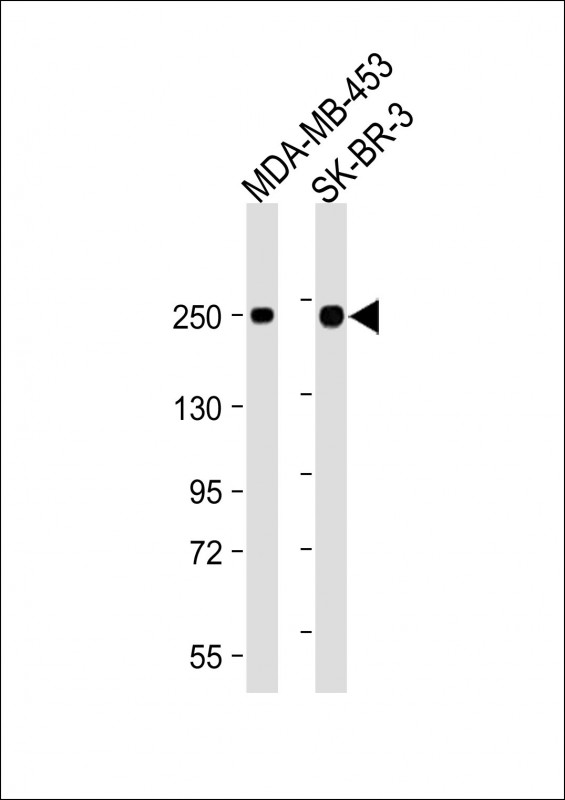
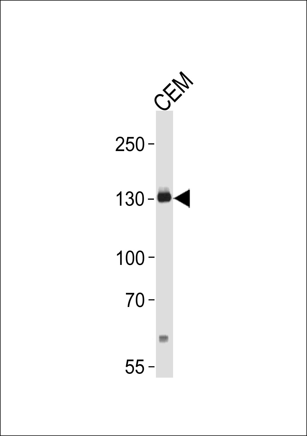
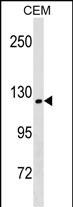
ERBB2 Antibody (C-term T1172/S1174)
Affinity Purified Rabbit Polyclonal Antibody (Pab)
- 产品详情
- 实验流程
- 背景知识
Application
| WB, E |
|---|---|
| Primary Accession | P04626 |
| Other Accession | P06494, P70424, NP_001005862.1 |
| Reactivity | Human, Mouse |
| Predicted | Mouse, Rat |
| Host | Rabbit |
| Clonality | Polyclonal |
| Isotype | Rabbit IgG |
| Calculated MW | 137910 Da |
| Antigen Region | 1151-1179 aa |
| Gene ID | 2064 |
|---|---|
| Other Names | Receptor tyrosine-protein kinase erbB-2, Metastatic lymph node gene 19 protein, MLN 19, Proto-oncogene Neu, Proto-oncogene c-ErbB-2, Tyrosine kinase-type cell surface receptor HER2, p185erbB2, CD340, ERBB2, HER2, MLN19, NEU, NGL |
| Target/Specificity | This ERBB2 antibody is generated from rabbits immunized with a KLH conjugated synthetic peptide between 1151-1179 amino acids from the C-terminal region of human ERBB2. |
| Dilution | WB~~1:1000 E~~Use at an assay dependent concentration. |
| Format | Purified polyclonal antibody supplied in PBS with 0.09% (W/V) sodium azide. This antibody is purified through a protein A column, followed by peptide affinity purification. |
| Storage | Maintain refrigerated at 2-8°C for up to 2 weeks. For long term storage store at -20°C in small aliquots to prevent freeze-thaw cycles. |
| Precautions | ERBB2 Antibody (C-term T1172/S1174) is for research use only and not for use in diagnostic or therapeutic procedures. |
| Name | ERBB2 |
|---|---|
| Synonyms | HER2, MLN19, NEU, NGL |
| Function | Protein tyrosine kinase that is part of several cell surface receptor complexes, but that apparently needs a coreceptor for ligand binding. Essential component of a neuregulin-receptor complex, although neuregulins do not interact with it alone. GP30 is a potential ligand for this receptor. Regulates outgrowth and stabilization of peripheral microtubules (MTs). Upon ERBB2 activation, the MEMO1-RHOA-DIAPH1 signaling pathway elicits the phosphorylation and thus the inhibition of GSK3B at cell membrane. This prevents the phosphorylation of APC and CLASP2, allowing its association with the cell membrane. In turn, membrane-bound APC allows the localization of MACF1 to the cell membrane, which is required for microtubule capture and stabilization. |
| Cellular Location | Cell membrane; Single-pass type I membrane protein. Cell projection, ruffle membrane; Single-pass type I membrane protein. Note=Internalized from the cell membrane in response to EGF stimulation. [Isoform 2]: Cytoplasm. Nucleus. |
| Tissue Location | Expressed in a variety of tumor tissues including primary breast tumors and tumors from small bowel, esophagus, kidney and mouth. |
For Research Use Only. Not For Use In Diagnostic Procedures.
Provided below are standard protocols that you may find useful for product applications.
BACKGROUND
This gene encodes a member of the epidermal growth factor (EGF) receptor family of receptor tyrosine kinases. This protein has no ligand binding domain of its own and therefore cannot bind growth factors. However, it does bind tightly to other ligand-bound EGF receptor family members to form a heterodimer, stabilizing ligand binding and enhancing kinase-mediated activation of downstream signalling pathways, such as those involving mitogen-activated protein kinase and phosphatidylinositol-3 kinase. Allelic variations at amino acid positions 654 and 655 of isoform a (positions 624 and 625 of isoform b) have been reported, with the most common allele, Ile654/Ile655, shown here. Amplification and/or overexpression of this gene has been reported in numerous cancers, including breast and ovarian tumors. Alternative splicing results in several additional transcript variants, some encoding different isoforms and others that have not been fully characterized.
REFERENCES
Geradts, J., et al. Cancer Invest. 28(9):969-977(2010)
Zaoui, K., et al. Proc. Natl. Acad. Sci. U.S.A. 107(43):18517-18522(2010)
Oliveras, G., et al. Ann. N. Y. Acad. Sci. 1210, 86-92 (2010) :
Han, J.S., et al. Anticancer Res. 30(9):3407-3412(2010)
Stackievicz, R., et al. Isr. Med. Assoc. J. 12(5):290-295(2010)
终于等到您。ABCEPTA(百远生物)抗体产品。
点击下方“我要评价 ”按钮提交您的反馈信息,您的反馈和评价是我们最宝贵的财富之一,
我们将在1-3个工作日内处理您的反馈信息。
如有疑问,联系:0512-88856768 tech-china@abcepta.com.


