


 癌症的基本特征包括细胞增殖、血管生成、迁移、凋亡逃避机制和细胞永生等。找到癌症发生过程中这些通路的关键标记物和对应的抗体用于检测至关重要。
 为您推荐一个泛素化位点预测神器——泛素化分析工具,可以为您的蛋白的泛素化位点作出预测和评分。
 细胞自噬受体图形绘图工具为你的蛋白的细胞受体结合位点作出预测和评分,识别结合到自噬通路中的蛋白是非常重要的,便于让我们理解自噬在正常生理、病理过程中的作用,如发育、细胞分化、神经退化性疾病、压力条件下、感染和癌症。
RNF128 Antibody (Center)
Affinity Purified Rabbit Polyclonal Antibody (Pab)
- 产品详情
- 实验流程
- 背景知识
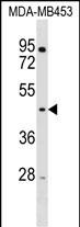
Application
| WB, E |
|---|---|
| Primary Accession | Q8TEB7 |
| Other Accession | Q29RU0, NP_078815.3 |
| Reactivity | Human |
| Predicted | Bovine |
| Host | Rabbit |
| Clonality | Polyclonal |
| Isotype | Rabbit IgG |
| Antigen Region | 115-143 aa |
| Other Names | E3 ubiquitin-protein ligase RNF128, 632-, Gene related to anergy in lymphocytes protein, GRAIL, RING finger protein 128, RNF128 |
|---|---|
| Target/Specificity | This RNF128 antibody is generated from rabbits immunized with a KLH conjugated synthetic peptide between 115-143 amino acids from the Central region of human RNF128. |
| Dilution | WB~~1:1000 E~~Use at an assay dependent concentration. |
| Format | Purified polyclonal antibody supplied in PBS with 0.09% (W/V) sodium azide. This antibody is purified through a protein A column, followed by peptide affinity purification. |
| Storage | Maintain refrigerated at 2-8°C for up to 2 weeks. For long term storage store at -20°C in small aliquots to prevent freeze-thaw cycles. |
| Precautions | RNF128 Antibody (Center) is for research use only and not for use in diagnostic or therapeutic procedures. |
For Research Use Only. Not For Use In Diagnostic Procedures.
Provided below are standard protocols that you may find useful for product applications.
BACKGROUND
The protein encoded by this gene is a type I transmembrane protein that localizes to the endocytic pathway. This protein contains a RING zinc-finger motif and has been shown to possess E3 ubiquitin ligase activity. Expression of this gene in retrovirally transduced T cell hybridoma significantly inhibits activation-induced IL2 and IL4 cytokine production. Induced expression of this gene was observed in anergic CD4(+) T cells, which suggested a role in the induction of anergic phenotype. Alternatively spliced transcript variants encoding distinct isoforms have been reported.
REFERENCES
Su, L.L., et al. J. Immunol. 183(1):438-444(2009)
Lin, J.T., et al. J. Immunol. 182(10):5919-5928(2009)
Lineberry, N., et al. J. Biol. Chem. 283(42):28497-28505(2008)
Egawa, S., et al. Am. J. Physiol. Gastrointest. Liver Physiol. 295 (1), G163-G169 (2008) :
Kostianovsky, A.M., et al. J. Immunol. 178(10):6158-6163(2007)
终于等到您。ABCEPTA(百远生物)抗体产品。
点击下方“我要评价 ”按钮提交您的反馈信息,您的反馈和评价是我们最宝贵的财富之一,
我们将在1-3个工作日内处理您的反馈信息。
如有疑问,联系:0512-88856768 tech-china@abcepta.com.


