


 癌症的基本特征包括细胞增殖、血管生成、迁移、凋亡逃避机制和细胞永生等。找到癌症发生过程中这些通路的关键标记物和对应的抗体用于检测至关重要。
 为您推荐一个泛素化位点预测神器——泛素化分析工具,可以为您的蛋白的泛素化位点作出预测和评分。
 细胞自噬受体图形绘图工具为你的蛋白的细胞受体结合位点作出预测和评分,识别结合到自噬通路中的蛋白是非常重要的,便于让我们理解自噬在正常生理、病理过程中的作用,如发育、细胞分化、神经退化性疾病、压力条件下、感染和癌症。
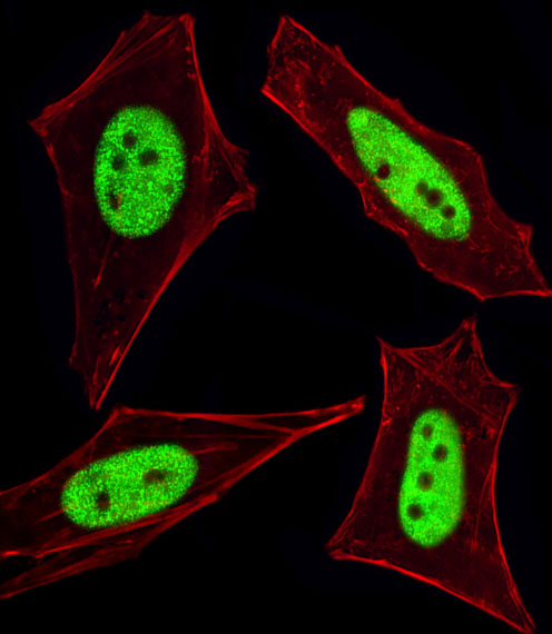
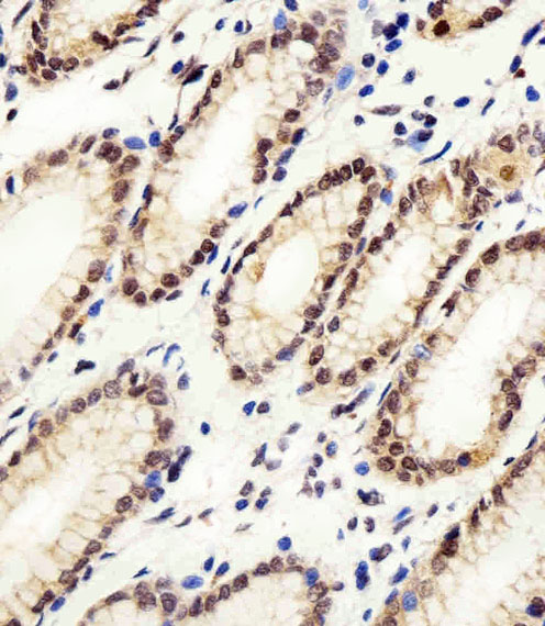
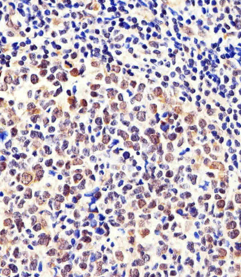
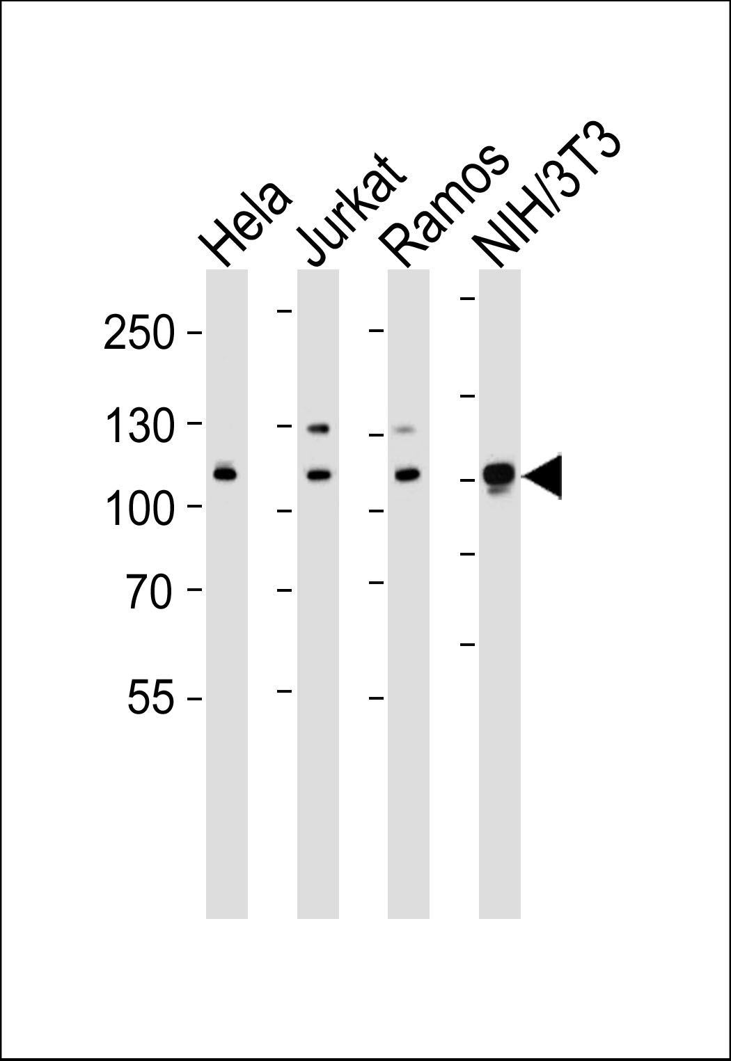
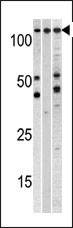
Cellular Apoptosis Susceptibility Antibody (C-term)
Purified Rabbit Polyclonal Antibody (Pab)
- 产品详情
- 文献引用 : 3
- 实验流程
- 背景知识
Application
| WB, IF, IHC-P, E |
|---|---|
| Primary Accession | P55060 |
| Other Accession | Q9ERK4, Q7SZC2, A5D785 |
| Reactivity | Human, Mouse |
| Predicted | Bovine, Zebrafish |
| Host | Rabbit |
| Clonality | Polyclonal |
| Isotype | Rabbit IgG |
| Calculated MW | 110417 Da |
| Antigen Region | 55-84 aa |
| Gene ID | 1434 |
|---|---|
| Other Names | Exportin-2, Exp2, Cellular apoptosis susceptibility protein, Chromosome segregation 1-like protein, Importin-alpha re-exporter, CSE1L, CAS, XPO2 |
| Target/Specificity | This Cellular Apoptosis Susceptibility antibody is generated from rabbits immunized with a KLH conjugated synthetic peptide between 55-84 amino acids from the N-terminal region of human Cellular Apoptosis Susceptibility. |
| Dilution | WB~~1:1000 IF~~1:100 IHC-P~~1:100 E~~Use at an assay dependent concentration. |
| Format | Purified polyclonal antibody supplied in PBS with 0.09% (W/V) sodium azide. This antibody is prepared by Saturated Ammonium Sulfate (SAS) precipitation followed by dialysis against PBS. |
| Storage | Maintain refrigerated at 2-8°C for up to 2 weeks. For long term storage store at -20°C in small aliquots to prevent freeze-thaw cycles. |
| Precautions | Cellular Apoptosis Susceptibility Antibody (C-term) is for research use only and not for use in diagnostic or therapeutic procedures. |
| Name | CSE1L |
|---|---|
| Synonyms | CAS {ECO:0000303|PubMed:7479798}, XPO2 |
| Function | Export receptor for importin-alpha. Mediates importin-alpha re-export from the nucleus to the cytoplasm after import substrates (cargos) have been released into the nucleoplasm. In the nucleus binds cooperatively to importin-alpha and to the GTPase Ran in its active GTP-bound form. Docking of this trimeric complex to the nuclear pore complex (NPC) is mediated through binding to nucleoporins. Upon transit of a nuclear export complex into the cytoplasm, disassembling of the complex and hydrolysis of Ran-GTP to Ran-GDP (induced by RANBP1 and RANGAP1, respectively) cause release of the importin-alpha from the export receptor. CSE1L/XPO2 then return to the nuclear compartment and mediate another round of transport. The directionality of nuclear export is thought to be conferred by an asymmetric distribution of the GTP- and GDP-bound forms of Ran between the cytoplasm and nucleus. |
| Cellular Location | Cytoplasm. Nucleus. Note=Shuttles between the nucleus and the cytoplasm. |
| Tissue Location | Detected in brain, placenta, ovary, testis and trachea (at protein level) (PubMed:10331944). Widely expressed (PubMed:10331944). Highly expressed in testis and in proliferating cells (PubMed:10331944, PubMed:7479798). |
For Research Use Only. Not For Use In Diagnostic Procedures.

Provided below are standard protocols that you may find useful for product applications.
BACKGROUND
Proteins that carry a nuclear localization signal (NLS) are transported into the nucleus by the importin-alpha/beta heterodimer. Importin-alpha binds the NLS, while importin-beta mediates translocation through the nuclear pore complex. After translocation, RanGTP binds importin-beta and displaces importin-alpha. Importin-alpha must then be returned to the cytoplasm, leaving the NLS protein behind. CSE1L binds strongly to NLS-free importin-alpha, and this binding is released in the cytoplasm by the combined action of RANBP1 and RANGAP1. In addition, CSE1L may play a role both in apoptosis and in cell proliferation.
REFERENCES
Goldberg, G.S., et al., J. Biol. Chem. 278(47):46533-46540 (2003).
Behrens, P., et al., Apoptosis 8(1):39-44 (2003).
Jiang, M.C., et al., Biochem. Biophys. Res. Commun. 294(4):900-905 (2002).
Wellmann, A., et al., Int. J. Mol. Med. 7(5):489-494 (2001).
Brinkmann, U., et al., Genomics 58(1):41-49 (1999).
终于等到您。ABCEPTA(百远生物)抗体产品。
点击下方“我要评价 ”按钮提交您的反馈信息,您的反馈和评价是我们最宝贵的财富之一,
我们将在1-3个工作日内处理您的反馈信息。
如有疑问,联系:0512-88856768 tech-china@abcepta.com.


