


 癌症的基本特征包括细胞增殖、血管生成、迁移、凋亡逃避机制和细胞永生等。找到癌症发生过程中这些通路的关键标记物和对应的抗体用于检测至关重要。
 为您推荐一个泛素化位点预测神器——泛素化分析工具,可以为您的蛋白的泛素化位点作出预测和评分。
 细胞自噬受体图形绘图工具为你的蛋白的细胞受体结合位点作出预测和评分,识别结合到自噬通路中的蛋白是非常重要的,便于让我们理解自噬在正常生理、病理过程中的作用,如发育、细胞分化、神经退化性疾病、压力条件下、感染和癌症。
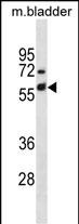
Mouse Krt6a Antibody(Center) 精选
Affinity Purified Rabbit Polyclonal Antibody (Pab)
- 产品详情
- 实验流程
- 背景知识
Application 
| WB, E |
|---|---|
| Primary Accession | P50446 |
| Other Accession | Q9Z331, NP_032502.3 |
| Reactivity | Mouse |
| Predicted | Rat, Bovine, Canine, Rabbit |
| Host | Rabbit |
| Clonality | Polyclonal |
| Isotype | Rabbit IgG |
| Calculated MW | 59335 Da |
| Antigen Region | 212-240 aa |
| Gene ID | 16687 |
|---|---|
| Other Names | Keratin, type II cytoskeletal 6A, Cytokeratin-6A, CK-6A, Keratin-6-alpha, mK6-alpha, Keratin-6A, K6A, Krt6a, Ker2, Krt2-6, Krt2-6a, Krt6 |
| Target/Specificity | This Mouse Krt6a antibody is generated from rabbits immunized with a KLH conjugated synthetic peptide between 212-240 amino acids from the Central region of mouse Krt6a. |
| Dilution | WB~~1:1000 E~~Use at an assay dependent concentration. |
| Format | Purified polyclonal antibody supplied in PBS with 0.09% (W/V) sodium azide. This antibody is purified through a protein A column, followed by peptide affinity purification. |
| Storage | Maintain refrigerated at 2-8°C for up to 2 weeks. For long term storage store at -20°C in small aliquots to prevent freeze-thaw cycles. |
| Precautions | Mouse Krt6a Antibody(Center) is for research use only and not for use in diagnostic or therapeutic procedures. |
For Research Use Only. Not For Use In Diagnostic Procedures.
| Name | Krt6a {ECO:0000312|MGI:MGI:1100845} |
|---|---|
| Function | Structural component of intermediate filaments in epithelial keratinocytes. Forms heteropolymers with the type I keratins KRT16 and KRT17, assembling keratin intermediate filament networks that contribute to the structural integrity and stress resilience of stratified epithelia, particularly in epidermis, nail bed and oral mucosa. Rapidly induced in wound-edge keratinocytes and participates in the reorganization of the cytoskeleton during re-epithelialization (PubMed:10866680, PubMed:8636216). Negatively regulates collective keratinocyte migration by stabilizing myosin IIA/MYH2 and desmoplakin, thereby altering cell-cell and cell-matrix adhesion and reducing the speed and directionality of epithelial sheet movement during wound repair (PubMed:30389720). Also limits epithelial migration by inhibiting the activity of SRC during wound repair (PubMed:22529101). Required for normal plantar and nail unit integrity (By similarity). |
| Cellular Location | Cytoplasm. |
| Tissue Location | Predominates in the adult trunk skin, tongue, trachea/esophagus and eye. In adult skin, localization is restricted to hair follicles, where it is localized predominantly in the outer root sheath. |
Provided below are standard protocols that you may find useful for product applications.
BACKGROUND
Keratins 6 and 16 are expressed in keratinocytes, which are undergoing rapid turnover in the suprabasal region (also known as hyperproliferation related keratins). Keratin 6 is found in hair follicles, neck squamous cell carcinomas, suprabasal cells of a variety of internal stratified epithelia, in epidermis, in both normal and hyperproliferative situations. Epidermal injury results in activation of keratinocytes which express CK6 and CK16. CK6 is strongly expressed in about 75% of head and neck squamous cell carcinomas. Expression of CK6 is particularly associated with differentiation. There are at least six isoforms of human type II keratin 6 (K6), K6A being the most abundant representing about 77% of all forms found in epithelia.
REFERENCES
Sehic, A., et al. Eur. J. Oral Sci. 117(2):93-104(2009)
Chen, J., et al. J. Invest. Dermatol. 128(2):270-279(2008)
Cui, C.Y., et al. Hum. Mol. Genet. 16(21):2583-2590(2007)
Gu, L.H., et al. J. Invest. Dermatol. 127(5):1061-1073(2007)
Kurek, D., et al. Development 134(2):261-272(2007)

终于等到您。ABCEPTA(百远生物)抗体产品。
点击下方“我要评价 ”按钮提交您的反馈信息,您的反馈和评价是我们最宝贵的财富之一,
我们将在1-3个工作日内处理您的反馈信息。
如有疑问,联系:0512-88856768 tech-china@abcepta.com.


