


 癌症的基本特征包括细胞增殖、血管生成、迁移、凋亡逃避机制和细胞永生等。找到癌症发生过程中这些通路的关键标记物和对应的抗体用于检测至关重要。
 为您推荐一个泛素化位点预测神器——泛素化分析工具,可以为您的蛋白的泛素化位点作出预测和评分。
 细胞自噬受体图形绘图工具为你的蛋白的细胞受体结合位点作出预测和评分,识别结合到自噬通路中的蛋白是非常重要的,便于让我们理解自噬在正常生理、病理过程中的作用,如发育、细胞分化、神经退化性疾病、压力条件下、感染和癌症。
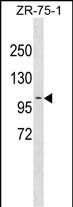
PDCD6IP Antibody (Center)
Affinity Purified Rabbit Polyclonal Antibody (Pab)
- 产品详情
- 实验流程
- 背景知识
Application
| WB, E |
|---|---|
| Primary Accession | Q8WUM4 |
| Other Accession | NP_037506.2 |
| Reactivity | Human |
| Host | Rabbit |
| Clonality | Polyclonal |
| Isotype | Rabbit IgG |
| Calculated MW | 96023 Da |
| Antigen Region | 541-570 aa |
| Gene ID | 10015 |
|---|---|
| Other Names | Programmed cell death 6-interacting protein, PDCD6-interacting protein, ALG-2-interacting protein 1, ALG-2-interacting protein X, Hp95, PDCD6IP, AIP1, ALIX, KIAA1375 |
| Target/Specificity | This PDCD6IP antibody is generated from rabbits immunized with a KLH conjugated synthetic peptide between 541-570 amino acids from the Central region of human PDCD6IP. |
| Dilution | WB~~1:1000 E~~Use at an assay dependent concentration. |
| Format | Purified polyclonal antibody supplied in PBS with 0.09% (W/V) sodium azide. This antibody is purified through a protein A column, followed by peptide affinity purification. |
| Storage | Maintain refrigerated at 2-8°C for up to 2 weeks. For long term storage store at -20°C in small aliquots to prevent freeze-thaw cycles. |
| Precautions | PDCD6IP Antibody (Center) is for research use only and not for use in diagnostic or therapeutic procedures. |
| Name | PDCD6IP (HGNC:8766) |
|---|---|
| Synonyms | AIP1, ALIX, KIAA1375 |
| Function | Multifunctional protein involved in endocytosis, multivesicular body biogenesis, membrane repair, cytokinesis, apoptosis and maintenance of tight junction integrity. Class E VPS protein involved in concentration and sorting of cargo proteins of the multivesicular body (MVB) for incorporation into intralumenal vesicles (ILVs) that are generated by invagination and scission from the limiting membrane of the endosome. Binds to the phospholipid lysobisphosphatidic acid (LBPA) which is abundant in MVBs internal membranes. The MVB pathway requires the sequential function of ESCRT-O, -I,-II and -III complexes (PubMed:14739459). The ESCRT machinery also functions in topologically equivalent membrane fission events, such as the terminal stages of cytokinesis (PubMed:17556548, PubMed:17853893). Adapter for a subset of ESCRT-III proteins, such as CHMP4, to function at distinct membranes. Required for completion of cytokinesis (PubMed:17556548, PubMed:17853893, PubMed:18641129). May play a role in the regulation of both apoptosis and cell proliferation. Regulates exosome biogenesis in concert with SDC1/4 and SDCBP (PubMed:22660413). By interacting with F-actin, PARD3 and TJP1 secures the proper assembly and positioning of actomyosin-tight junction complex at the apical sides of adjacent epithelial cells that defines a spatial membrane domain essential for the maintenance of epithelial cell polarity and barrier (By similarity). |
| Cellular Location | Cytoplasm, cytosol {ECO:0000250|UniProtKB:Q9QZA2}. Melanosome. Cytoplasm, cytoskeleton, microtubule organizing center, centrosome. Secreted, extracellular exosome. Cell junction, tight junction {ECO:0000250|UniProtKB:Q9WU78}. Midbody, Midbody ring Note=Identified by mass spectrometry in melanosome fractions from stage I to stage IV. Colocalized with CEP55 at centrosomes of non-dividing cells. Component of the actomyosin-tight junction complex (By similarity). PDCD6IP targeting to the midbody requires the interaction with CEP55 (PubMed:18641129). {ECO:0000250|UniProtKB:Q9QZA2, ECO:0000250|UniProtKB:Q9WU78, ECO:0000269|PubMed:17081065, ECO:0000269|PubMed:17556548, ECO:0000269|PubMed:17853893, ECO:0000269|PubMed:18641129} |
For Research Use Only. Not For Use In Diagnostic Procedures.
Provided below are standard protocols that you may find useful for product applications.
BACKGROUND
This gene encodes a protein thought to participate in programmed cell death. Studies using mouse cells have shown that overexpression of this protein can block apoptosis. In addition, the product of this gene binds to the product of the PDCD6 gene, a protein required for apoptosis, in a calcium-dependent manner. This gene product also binds to endophilins, proteins that regulate membrane shape during endocytosis. Overexpression of this gene product and endophilins results in cytoplasmic vacuolization, which may be partly responsible for the protection against cell death. Several alternatively spliced transcript variants encoding different isoforms have been found for this gene. [provided by RefSeq].
REFERENCES
Shi, X., et al. Biochem. J. 431(1):93-102(2010)
Irie, T., et al. Virology 405(2):334-341(2010)
Sette, P., et al. J. Virol. 84(16):8181-8192(2010)
Rose, J.E., et al. Mol. Med. 16 (7-8), 247-253 (2010) :
Inuzuka, T., et al. BMC Struct. Biol. 10, 25 (2010) :
终于等到您。ABCEPTA(百远生物)抗体产品。
点击下方“我要评价 ”按钮提交您的反馈信息,您的反馈和评价是我们最宝贵的财富之一,
我们将在1-3个工作日内处理您的反馈信息。
如有疑问,联系:0512-88856768 tech-china@abcepta.com.


