


 癌症的基本特征包括细胞增殖、血管生成、迁移、凋亡逃避机制和细胞永生等。找到癌症发生过程中这些通路的关键标记物和对应的抗体用于检测至关重要。
 为您推荐一个泛素化位点预测神器——泛素化分析工具,可以为您的蛋白的泛素化位点作出预测和评分。
 细胞自噬受体图形绘图工具为你的蛋白的细胞受体结合位点作出预测和评分,识别结合到自噬通路中的蛋白是非常重要的,便于让我们理解自噬在正常生理、病理过程中的作用,如发育、细胞分化、神经退化性疾病、压力条件下、感染和癌症。
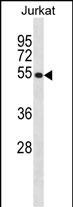
CREB3 Antibody (N-term)
Affinity Purified Rabbit Polyclonal Antibody (Pab)
- 产品详情
- 实验流程
- 背景知识
Application
| WB, E |
|---|---|
| Primary Accession | O43889 |
| Other Accession | NP_006359.3 |
| Reactivity | Human |
| Host | Rabbit |
| Clonality | Polyclonal |
| Isotype | Rabbit IgG |
| Calculated MW | 41379 Da |
| Antigen Region | 66-94 aa |
| Gene ID | 10488 |
|---|---|
| Other Names | Cyclic AMP-responsive element-binding protein 3, CREB-3, cAMP-responsive element-binding protein 3, Leucine zipper protein, Luman, Transcription factor LZIP-alpha, Processed cyclic AMP-responsive element-binding protein 3, N-terminal Luman, Transcriptionally active form, CREB3, LZIP |
| Target/Specificity | This CREB3 antibody is generated from rabbits immunized with a KLH conjugated synthetic peptide between 66-94 amino acids from the N-terminal region of human CREB3. |
| Dilution | WB~~1:1000 E~~Use at an assay dependent concentration. |
| Format | Purified polyclonal antibody supplied in PBS with 0.09% (W/V) sodium azide. This antibody is purified through a protein A column, followed by peptide affinity purification. |
| Storage | Maintain refrigerated at 2-8°C for up to 2 weeks. For long term storage store at -20°C in small aliquots to prevent freeze-thaw cycles. |
| Precautions | CREB3 Antibody (N-term) is for research use only and not for use in diagnostic or therapeutic procedures. |
| Name | CREB3 |
|---|---|
| Synonyms | LZIP |
| Function | Endoplasmic reticulum (ER)-bound sequence-specific transcription factor that directly binds DNA and activates transcription (PubMed:10984507, PubMed:15845366, PubMed:16940180, PubMed:19779205, PubMed:9271389). Plays a role in the unfolded protein response (UPR), promoting cell survival versus ER stress-induced apoptotic cell death (PubMed:15845366, PubMed:16940180). Also involved in cell proliferation, migration and differentiation, tumor suppression and inflammatory gene expression. Acts as a positive regulator of LKN- 1/CCL15-induced chemotaxis signaling of leukocyte cell migration (PubMed:15001559, PubMed:17296613, PubMed:19779205). Associates with chromatin to the HERPUD1 promoter (PubMed:16940180). Also induces transcriptional activation of chemokine receptors (PubMed:17296613, PubMed:18587271). |
| Cellular Location | [Isoform 1]: Endoplasmic reticulum membrane; Single-pass type II membrane protein {ECO:0000255, ECO:0000269|PubMed:12138176}. Golgi apparatus. Note=Colocalizes with HCFC1 in neuronal cell bodies of the trigeminal ganglia (PubMed:10623756). Colocalizes with DCSTAMP in the ER membrane of immature dendritic cell (DC) (PubMed:20546900). Colocalizes with CANX, CCR1, HCFC1 in the ER membrane (PubMed:10623756). [Isoform 2]: Nucleus. Cytoplasm Note=Predominantly in the nucleus (PubMed:19779205). Not associated with membranes (PubMed:19779205). |
| Tissue Location | Ubiquitously expressed (PubMed:19779205, PubMed:9271389). Expressed in dendritic cells (DC). Weakly expressed in monocytes (at protein level) (PubMed:20546900) |
For Research Use Only. Not For Use In Diagnostic Procedures.
Provided below are standard protocols that you may find useful for product applications.
BACKGROUND
This gene encodes a transcription factor that is a member of the leucine zipper family of DNA binding proteins. This protein binds to the cAMP-response element and regulates cell proliferation. The protein interacts with host cell factor C1, which also associates with the herpes simplex virus (HSV) protein VP16 that induces transcription of HSV immediate-early genes. This protein and VP16 both bind to the same site on host cell factor C1. It is thought that the interaction between this protein and host cell factor C1 plays a role in the establishment of latency during HSV infection. This protein also plays a role in leukocyte migration, tumor suppression, and endoplasmic reticulum stress-associated protein degradation. Additional transcript variants have been identified, but their biological validity has not been determined.
REFERENCES
Kim, H.C., et al. Cell. Mol. Life Sci. 67(20):3499-3510(2010)
Eleveld-Trancikova, D., et al. Mol. Immunol. 47 (11-12), 1963-1973 (2010) :
Kang, H., et al. Mol. Endocrinol. 23(11):1746-1757(2009)
Mamdani, F., et al. Am. J. Med. Genet. B Neuropsychiatr. Genet. 147B (4), 500-504 (2008) :
Audas, T.E., et al. Mol. Cell. Biol. 28(12):3952-3966(2008)
终于等到您。ABCEPTA(百远生物)抗体产品。
点击下方“我要评价 ”按钮提交您的反馈信息,您的反馈和评价是我们最宝贵的财富之一,
我们将在1-3个工作日内处理您的反馈信息。
如有疑问,联系:0512-88856768 tech-china@abcepta.com.


