


 癌症的基本特征包括细胞增殖、血管生成、迁移、凋亡逃避机制和细胞永生等。找到癌症发生过程中这些通路的关键标记物和对应的抗体用于检测至关重要。
 为您推荐一个泛素化位点预测神器——泛素化分析工具,可以为您的蛋白的泛素化位点作出预测和评分。
 细胞自噬受体图形绘图工具为你的蛋白的细胞受体结合位点作出预测和评分,识别结合到自噬通路中的蛋白是非常重要的,便于让我们理解自噬在正常生理、病理过程中的作用,如发育、细胞分化、神经退化性疾病、压力条件下、感染和癌症。
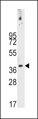
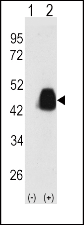
PDX1 Antibody (T11)
Affinity Purified Rabbit Polyclonal Antibody (Pab)
_-_SY5Y.jpg)
- 产品详情
- 实验流程
- 背景知识
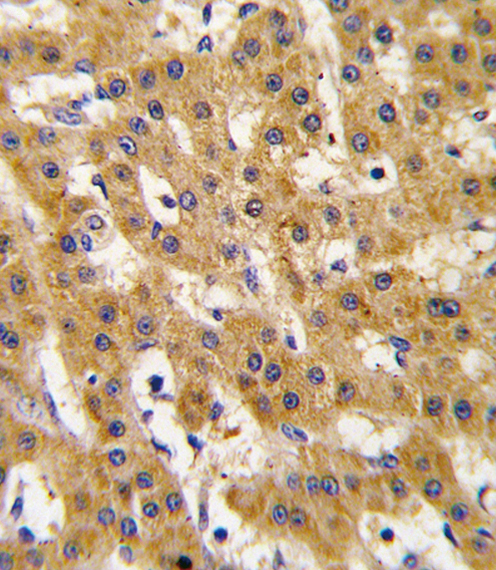
Application
| WB, IF, IHC-P, E |
|---|---|
| Primary Accession | O00330 |
| Other Accession | P52947, P52946, P52945 |
| Reactivity | Human, Rat, Mouse |
| Predicted | Mouse, Rat |
| Host | Rabbit |
| Clonality | Polyclonal |
| Isotype | Rabbit IgG |
| Calculated MW | 54122 Da |
| Antigen Region | 1-30 aa |
| Gene ID | 8050 |
|---|---|
| Other Names | Pyruvate dehydrogenase protein X component, mitochondrial, Dihydrolipoamide dehydrogenase-binding protein of pyruvate dehydrogenase complex, E3-binding protein, E3BP, Lipoyl-containing pyruvate dehydrogenase complex component X, proX, PDHX, PDX1 |
| Target/Specificity | This PDX1 antibody is generated from rabbits immunized with a KLH conjugated synthetic peptide between 1-30 amino acids from human PDX1. |
| Dilution | WB~~1:1000 IF~~1:100 IHC-P~~1:100~500 E~~Use at an assay dependent concentration. |
| Format | Purified polyclonal antibody supplied in PBS with 0.09% (W/V) sodium azide. This antibody is purified through a protein A column, followed by peptide affinity purification. |
| Storage | Maintain refrigerated at 2-8°C for up to 2 weeks. For long term storage store at -20°C in small aliquots to prevent freeze-thaw cycles. |
| Precautions | PDX1 Antibody (T11) is for research use only and not for use in diagnostic or therapeutic procedures. |
| Name | PDHX |
|---|---|
| Synonyms | PDX1 |
| Function | Required for anchoring dihydrolipoamide dehydrogenase (E3) to the dihydrolipoamide transacetylase (E2) core of the pyruvate dehydrogenase complexes of eukaryotes. This specific binding is essential for a functional PDH complex. |
| Cellular Location | Mitochondrion matrix. |
For Research Use Only. Not For Use In Diagnostic Procedures.
Provided below are standard protocols that you may find useful for product applications.
BACKGROUND
PDX1, located in the mitochondrial matrix, is required for anchoring dihydrolipoamide dehydrogenase (E3) to the dihydrolipoamide transacetylase (E2) core of the pyruvate dehydrogenase complexes of eukaryotes. This specific binding is essential for a functional PDH complex. Eukaryotic pyruvate dehydrogenase complexes are organized about a core consisting of the oligomeric dihydrolipoamide acetyl-transferase, around which are arranged multiple copies of pyruvate dehydrogenase, dihydrolipoamide dehydrogenase and protein X bound by noncovalent bonds. Defects in PDHX are a cause of lacticacidemia. PDX1 belongs to the 2-oxoacid dehydrogenase family and contains 1 lipoyl-binding domain.
REFERENCES
References for protein:
1.Ling, M., et al., Hum. Mol. Genet. 7(3):501-505 (1998).
2.Aral, B., et al., Am. J. Hum. Genet. 61(6):1318-1326 (1997).
3.Harris, R.A., et al., J. Biol. Chem. 272(32):19746-19751 (1997).
4.Yu, W., et al., Genome Res. 7(4):353-358 (1997).
References for SY5Y (SH-SY5Y; ATCC#CRL-2266): 1. Ross RA, et al. Coordinate morphological and biochemical interconversion of human neuroblastoma cells. J. Natl. Cancer Inst. 71: 741-749, 1983. [PubMed: 6137586]; 2. Biedler JL, et al. Multiple neurotransmitter synthesis by human neuroblastoma cell lines and clones. Cancer Res. 38: 3751-3757, 1978. [PubMed: 29704]
终于等到您。ABCEPTA(百远生物)抗体产品。
点击下方“我要评价 ”按钮提交您的反馈信息,您的反馈和评价是我们最宝贵的财富之一,
我们将在1-3个工作日内处理您的反馈信息。
如有疑问,联系:0512-88856768 tech-china@abcepta.com.


