


 癌症的基本特征包括细胞增殖、血管生成、迁移、凋亡逃避机制和细胞永生等。找到癌症发生过程中这些通路的关键标记物和对应的抗体用于检测至关重要。
 为您推荐一个泛素化位点预测神器——泛素化分析工具,可以为您的蛋白的泛素化位点作出预测和评分。
 细胞自噬受体图形绘图工具为你的蛋白的细胞受体结合位点作出预测和评分,识别结合到自噬通路中的蛋白是非常重要的,便于让我们理解自噬在正常生理、病理过程中的作用,如发育、细胞分化、神经退化性疾病、压力条件下、感染和癌症。
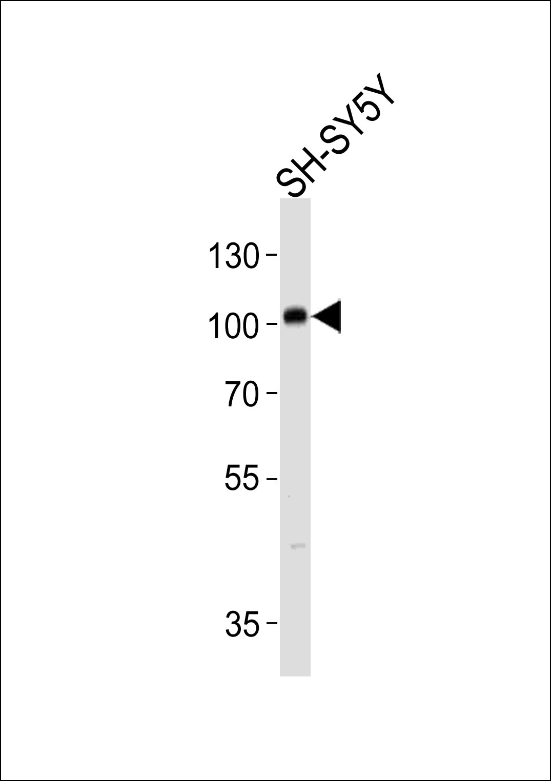
PROX1 Antibody (S197)
Affinity Purified Rabbit Polyclonal Antibody (Pab)
- 产品详情
- 实验流程
- 背景知识
Application
| WB, E |
|---|---|
| Primary Accession | Q92786 |
| Other Accession | P48437, NP_002754 |
| Reactivity | Human, Mouse |
| Predicted | Mouse |
| Host | Rabbit |
| Clonality | Polyclonal |
| Isotype | Rabbit IgG |
| Calculated MW | 83203 Da |
| Antigen Region | 175-206 aa |
| Gene ID | 5629 |
|---|---|
| Other Names | Prospero homeobox protein 1, Homeobox prospero-like protein PROX1, PROX-1, PROX1 |
| Target/Specificity | This PROX1 antibody is generated from rabbits immunized with a KLH conjugated synthetic peptide between 175-206 amino acids from human PROX1. |
| Dilution | WB~~1:1000 E~~Use at an assay dependent concentration. |
| Format | Purified polyclonal antibody supplied in PBS with 0.09% (W/V) sodium azide. This antibody is purified through a protein A column, followed by peptide affinity purification. |
| Storage | Maintain refrigerated at 2-8°C for up to 2 weeks. For long term storage store at -20°C in small aliquots to prevent freeze-thaw cycles. |
| Precautions | PROX1 Antibody (S197) is for research use only and not for use in diagnostic or therapeutic procedures. |
| Name | PROX1 |
|---|---|
| Function | Transcription factor involved in developmental processes such as cell fate determination, gene transcriptional regulation and progenitor cell regulation in a number of organs. Plays a critical role in embryonic development and functions as a key regulatory protein in neurogenesis and the development of the heart, eye lens, liver, pancreas and the lymphatic system. Involved in the regulation of the circadian rhythm. Represses: transcription of the retinoid-related orphan receptor RORG, transcriptional activator activity of RORA and RORG and the expression of RORA/G-target genes including core clock components: BMAL1, NPAS2 and CRY1 and metabolic genes: AVPR1A and ELOVL3. |
| Cellular Location | Nucleus {ECO:0000250|UniProtKB:P48437}. Note=RORG promotes its nuclear localization. {ECO:0000250|UniProtKB:P48437} |
| Tissue Location | Most actively expressed in the developing lens. Detected also in embryonic brain, lung, liver and kidney. In adult, it is more abundant in heart and liver than in brain, skeletal muscle, kidney and pancreas. |
For Research Use Only. Not For Use In Diagnostic Procedures.
Provided below are standard protocols that you may find useful for product applications.
BACKGROUND
The expression pattern of Prox1 suggests that it has a role in a variety of embryonic tissues, including lens. Prox mRNA is present in many different human tissues with lens demonstrating the highest level. Homozygous Prox1-null mice die at midgestation from multiple developmental defects, and a targeted effect on lens development has been reported. Prox1 inactivation caused abnormal cellular proliferation, downregulated expression of the cell cycle inhibitors Cdkn1b and Cdkn1c, misexpression of E-cadherin, and excessive apoptosis. Consequently, mutant lens cells failed to polarize and elongate properly, resulting in a hollow lens. Prox1 is expressed in a subpopulation of endothelial cells that by budding and sprouting give rise to the lymphatic system. Prox1 appears to be a specific and required regulator of the development of the lymphatic system. Prox1 also has been documented to be required for hepatocyte migration in the mouse. Loss of Prox1 results in a smaller liver with a reduced population of clustered hepatocytes. The homeodomain protein Prox1 regulates the egress of progenitor cells from the cell cycle in the embryonic mouse retina. Cells lacking Prox1 are less likely to stop dividing, and ectopic expression of Prox1 forces progenitor cells to exit the cell cycle. Prox1 acts as a key participant in progenitor-cell proliferation and cell-fate determination in the vertebrate retina.
REFERENCES
Nagai, H., et al., Genes Chromosomes Cancer 38(1):13-21 (2003).
Dyer, M.A., et al., Nat. Genet. 34(1):53-58 (2003).
Hong, Y.K., et al., Dev. Dyn. 225(3):351-357 (2002).
Petrova, T.V., et al., EMBO J. 21(17):4593-4599 (2002).
Mouta Carreira, C., et al., Cancer Res. 61(22):8079-8084 (2001).
终于等到您。ABCEPTA(百远生物)抗体产品。
点击下方“我要评价 ”按钮提交您的反馈信息,您的反馈和评价是我们最宝贵的财富之一,
我们将在1-3个工作日内处理您的反馈信息。
如有疑问,联系:0512-88856768 tech-china@abcepta.com.


