


 癌症的基本特征包括细胞增殖、血管生成、迁移、凋亡逃避机制和细胞永生等。找到癌症发生过程中这些通路的关键标记物和对应的抗体用于检测至关重要。
 为您推荐一个泛素化位点预测神器——泛素化分析工具,可以为您的蛋白的泛素化位点作出预测和评分。
 细胞自噬受体图形绘图工具为你的蛋白的细胞受体结合位点作出预测和评分,识别结合到自噬通路中的蛋白是非常重要的,便于让我们理解自噬在正常生理、病理过程中的作用,如发育、细胞分化、神经退化性疾病、压力条件下、感染和癌症。
PARP3 Antibody (N-term)
Affinity Purified Rabbit Polyclonal Antibody (Pab)
- 产品详情
- 实验流程
- 背景知识
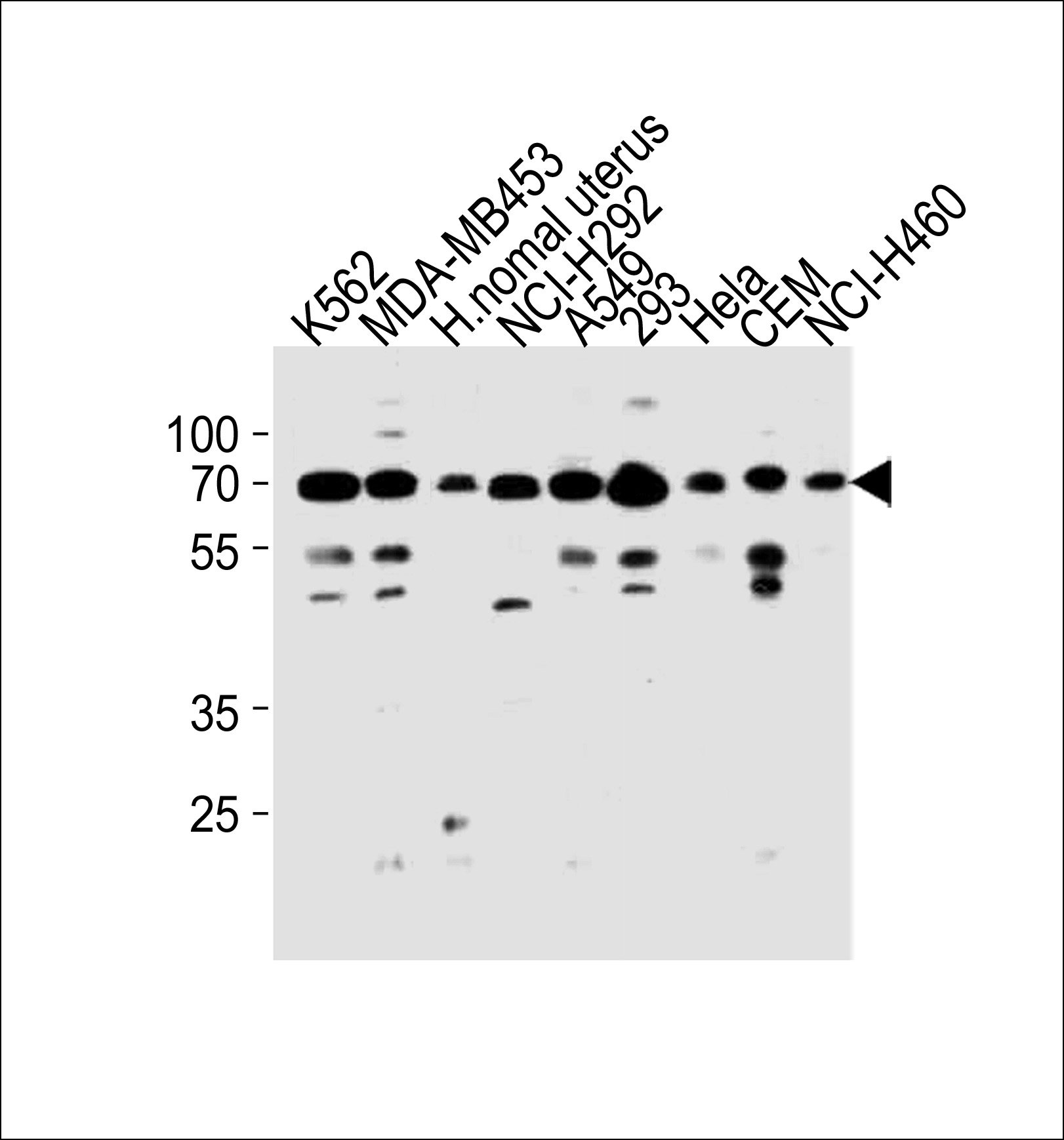
Application
| WB, E |
|---|---|
| Primary Accession | Q9Y6F1 |
| Reactivity | Human |
| Host | Rabbit |
| Clonality | Polyclonal |
| Isotype | Rabbit IgG |
| Calculated MW | 60089 Da |
| Antigen Region | 99-126 aa |
| Gene ID | 10039 |
|---|---|
| Other Names | Poly [ADP-ribose] polymerase 3, PARP-3, hPARP-3, ADP-ribosyltransferase diphtheria toxin-like 3, ARTD3, IRT1, NAD(+) ADP-ribosyltransferase 3, ADPRT-3, Poly[ADP-ribose] synthase 3, pADPRT-3, PARP3, ADPRT3, ADPRTL3 |
| Target/Specificity | This PARP3 antibody is generated from rabbits immunized with a KLH conjugated synthetic peptide between 99-126 amino acids from the N-terminal region of human PARP3. |
| Dilution | WB~~1:1000 E~~Use at an assay dependent concentration. |
| Format | Purified polyclonal antibody supplied in PBS with 0.09% (W/V) sodium azide. This antibody is purified through a protein A column, followed by peptide affinity purification. |
| Storage | Maintain refrigerated at 2-8°C for up to 2 weeks. For long term storage store at -20°C in small aliquots to prevent freeze-thaw cycles. |
| Precautions | PARP3 Antibody (N-term) is for research use only and not for use in diagnostic or therapeutic procedures. |
| Name | PARP3 {ECO:0000303|PubMed:10329013, ECO:0000312|HGNC:HGNC:273} |
|---|---|
| Function | Mono-ADP-ribosyltransferase that mediates mono-ADP- ribosylation of target proteins and plays a key role in the response to DNA damage (PubMed:16924674, PubMed:19354255, PubMed:20064938, PubMed:21211721, PubMed:21270334, PubMed:23742272, PubMed:24598253, PubMed:25043379, PubMed:28447610). Mediates mono-ADP-ribosylation of glutamate, aspartate or lysine residues on target proteins (PubMed:20064938, PubMed:25043379). In contrast to PARP1 and PARP2, it is not able to mediate poly-ADP-ribosylation (PubMed:25043379). Involved in DNA repair by mediating mono-ADP-ribosylation of a limited number of acceptor proteins involved in chromatin architecture and in DNA metabolism, such as histone H2B, XRCC5 and XRCC6 (PubMed:16924674, PubMed:24598253). ADP-ribosylation follows DNA damage and appears as an obligatory step in a detection/signaling pathway leading to the reparation of DNA strand breaks (PubMed:16924674, PubMed:21211721, PubMed:21270334). Involved in single-strand break repair by catalyzing mono-ADP-ribosylation of histone H2B on 'Glu-2' (H2BE2ADPr) of nucleosomes containing nicked DNA (PubMed:27530147). Cooperates with the XRCC5-XRCC6 (Ku80-Ku70) heterodimer to limit end-resection thereby promoting accurate NHEJ (PubMed:24598253). Suppresses G-quadruplex (G4) structures in response to DNA damage (PubMed:28447610). Associates with a number of DNA repair factors and is involved in the response to exogenous and endogenous DNA strand breaks (PubMed:16924674, PubMed:21211721, PubMed:21270334). Together with APLF, promotes the retention of the LIG4-XRCC4 complex on chromatin and accelerate DNA ligation during non-homologous end-joining (NHEJ) (PubMed:21211721). May link the DNA damage surveillance network to the mitotic fidelity checkpoint (PubMed:16924674). Acts as a negative regulator of immunoglobulin class switch recombination, probably by controlling the level of AICDA /AID on the chromatin (By similarity). In addition to proteins, also able to ADP-ribosylate DNA: mediates DNA mono-ADP- ribosylation of DNA strand break termini via covalent addition of a single ADP-ribose moiety to a 5'- or 3'-terminal phosphate residues in DNA containing multiple strand breaks (PubMed:29361132, PubMed:29520010). |
| Cellular Location | Nucleus. Chromosome. Cytoplasm, cytoskeleton, microtubule organizing center, centrosome. Cytoplasm, cytoskeleton, microtubule organizing center, centrosome, centriole. Note=Almost exclusively localized in the nucleus and appears in numerous small foci and a small number of larger foci whereas a centrosomal location has not been detected (PubMed:16924674). In response to DNA damage, localizes to sites of double-strand break (PubMed:21270334, PubMed:28447610). Also localizes to single-strand breaks (PubMed:27530147). Preferentially localized to the daughter centriole (PubMed:10329013). |
| Tissue Location | Widely expressed; the highest levels are in the kidney, skeletal muscle, liver, heart and spleen; also detected in pancreas, lung, placenta, brain, leukocytes, colon, small intestine, ovary, testis, prostate and thymus. |
For Research Use Only. Not For Use In Diagnostic Procedures.
Provided below are standard protocols that you may find useful for product applications.
BACKGROUND
Involved in the base excision repair (BER) pathway, by catalyzing the poly(ADP-ribosyl)ation of a limited number of acceptor proteins involved in chromatin architecture and in DNA metabolism. This modification follows DNA damages and appears as an obligatory step in a detection/signaling pathway leading to the reparation of DNA strand breaks. May link the DNA damage surveillance network to the mitotic fidelity checkpoint. Negatively influences the G1/S cell cycle progression without interfering with centrosome duplication. Binds DNA. May be involved in the regulation of PRC2 and PRC3 complex-dependent gene silencing.
终于等到您。ABCEPTA(百远生物)抗体产品。
点击下方“我要评价 ”按钮提交您的反馈信息,您的反馈和评价是我们最宝贵的财富之一,
我们将在1-3个工作日内处理您的反馈信息。
如有疑问,联系:0512-88856768 tech-china@abcepta.com.


