


 癌症的基本特征包括细胞增殖、血管生成、迁移、凋亡逃避机制和细胞永生等。找到癌症发生过程中这些通路的关键标记物和对应的抗体用于检测至关重要。
 为您推荐一个泛素化位点预测神器——泛素化分析工具,可以为您的蛋白的泛素化位点作出预测和评分。
 细胞自噬受体图形绘图工具为你的蛋白的细胞受体结合位点作出预测和评分,识别结合到自噬通路中的蛋白是非常重要的,便于让我们理解自噬在正常生理、病理过程中的作用,如发育、细胞分化、神经退化性疾病、压力条件下、感染和癌症。
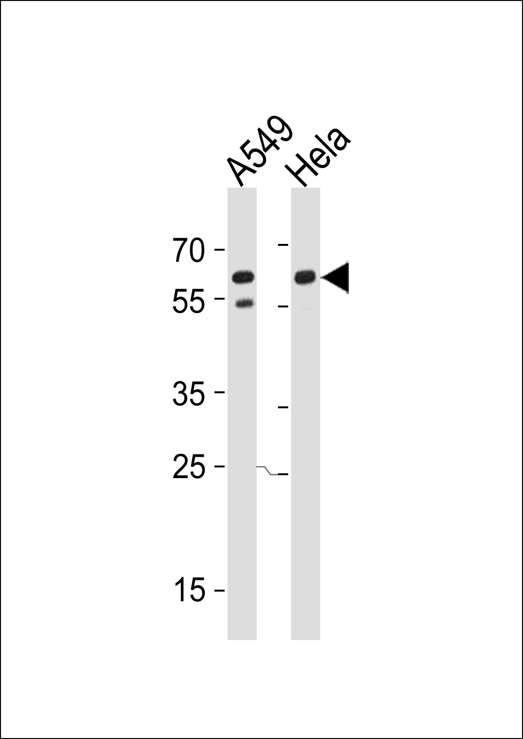
CD46 Antibody (Center Y354)
Affinity Purified Rabbit Polyclonal Antibody (Pab)
- 产品详情
- 实验流程
- 背景知识
Application
| WB, E |
|---|---|
| Primary Accession | P15529 |
| Reactivity | Human |
| Host | Rabbit |
| Clonality | Polyclonal |
| Isotype | Rabbit IgG |
| Antigen Region | 330-361 aa |
| Other Names | Membrane cofactor protein, TLX, Trophoblast leukocyte common antigen, CD46, CD46, MCP, MIC10 |
|---|---|
| Target/Specificity | This CD46 antibody is generated from rabbits immunized with a KLH conjugated synthetic peptide between 330-361 amino acids from the Central region of human CD46. |
| Dilution | WB~~1:1000 E~~Use at an assay dependent concentration. |
| Format | Purified polyclonal antibody supplied in PBS with 0.09% (W/V) sodium azide. This antibody is purified through a protein A column, followed by peptide affinity purification. |
| Storage | Maintain refrigerated at 2-8°C for up to 2 weeks. For long term storage store at -20°C in small aliquots to prevent freeze-thaw cycles. |
| Precautions | CD46 Antibody (Center Y354) is for research use only and not for use in diagnostic or therapeutic procedures. |
For Research Use Only. Not For Use In Diagnostic Procedures.
Provided below are standard protocols that you may find useful for product applications.
BACKGROUND
Acts as a cofactor for complement factor I, a serine protease which protects autologous cells against complement-mediated injury by cleaving C3b and C4b deposited on host tissue. May be involved in the fusion of the spermatozoa with the oocyte during fertilization. Also acts as a costimulatory factor for T-cells which induces the differentiation of CD4+ into T-regulatory 1 cells. T-regulatory 1 cells suppress immune responses by secreting interleukin-10, and therefore are thought to prevent autoimmunity. A number of viral and bacterial pathogens seem to exploit this property and directly induce an immunosuppressive phenotype in T-cells by binding to CD46.
REFERENCES
Riley R.C., et al. Mol. Reprod. Dev. 62:534-546(2002).
Lublin D.M., et al. J. Exp. Med. 168:181-194(1988).
Purcell D.F., et al. Immunogenetics 33:335-344(1991).
Post T.W., et al. J. Exp. Med. 174:93-102(1991).
Cervoni F., et al. Mol. Reprod. Dev. 34:107-113(1993).
终于等到您。ABCEPTA(百远生物)抗体产品。
点击下方“我要评价 ”按钮提交您的反馈信息,您的反馈和评价是我们最宝贵的财富之一,
我们将在1-3个工作日内处理您的反馈信息。
如有疑问,联系:0512-88856768 tech-china@abcepta.com.


