


 癌症的基本特征包括细胞增殖、血管生成、迁移、凋亡逃避机制和细胞永生等。找到癌症发生过程中这些通路的关键标记物和对应的抗体用于检测至关重要。
 为您推荐一个泛素化位点预测神器——泛素化分析工具,可以为您的蛋白的泛素化位点作出预测和评分。
 细胞自噬受体图形绘图工具为你的蛋白的细胞受体结合位点作出预测和评分,识别结合到自噬通路中的蛋白是非常重要的,便于让我们理解自噬在正常生理、病理过程中的作用,如发育、细胞分化、神经退化性疾病、压力条件下、感染和癌症。
MYH14 Antibody (Center)
Purified Rabbit Polyclonal Antibody (Pab)
- 产品详情
- 实验流程
- 背景知识
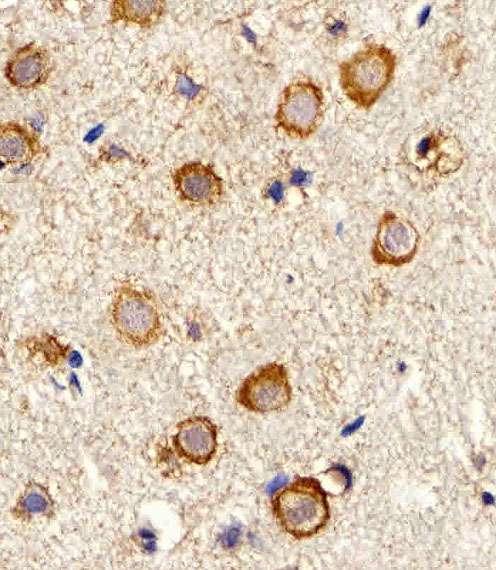
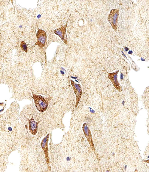
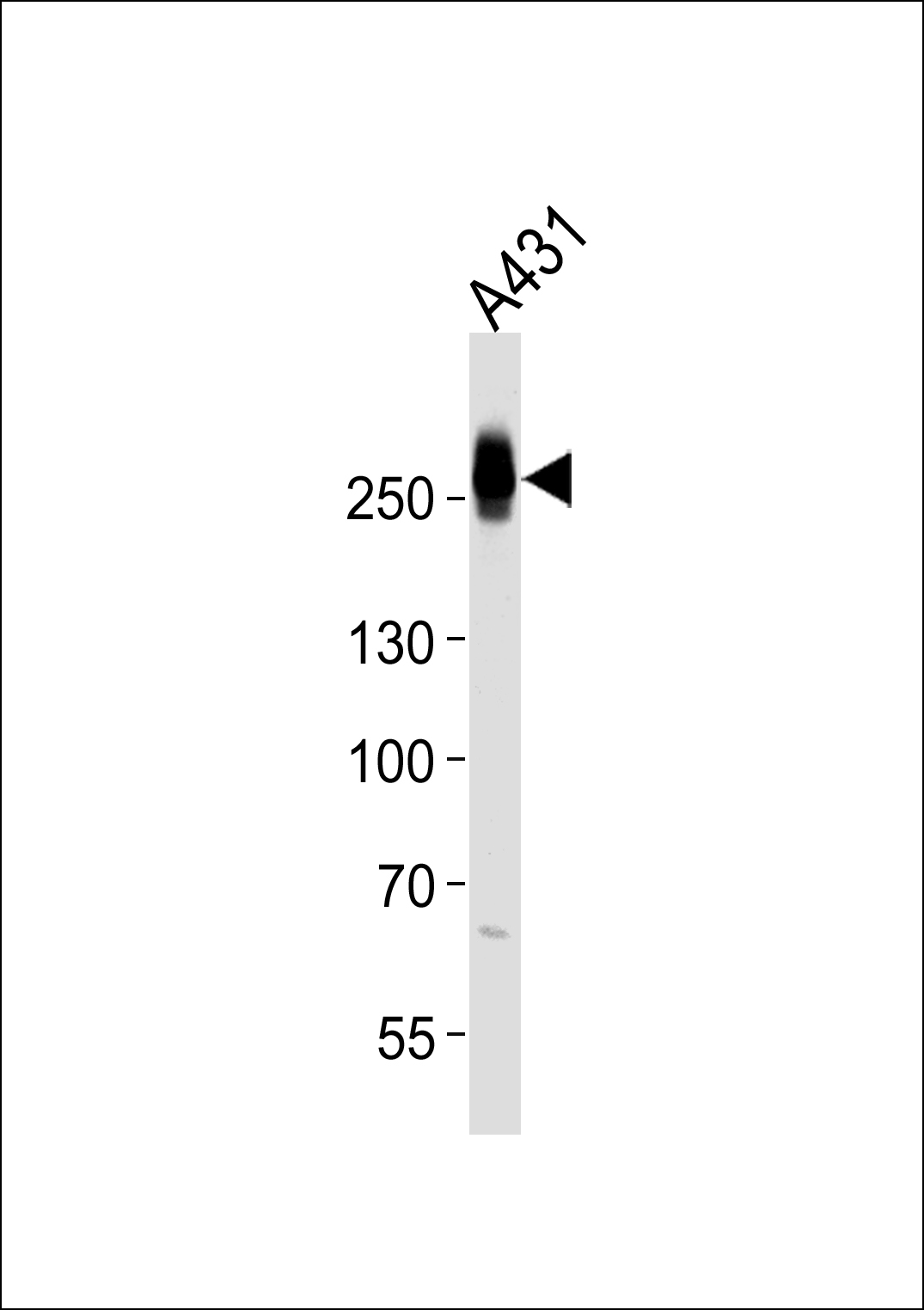
Application
| WB, IHC-P, E |
|---|---|
| Primary Accession | Q7Z406 |
| Reactivity | Human |
| Host | Rabbit |
| Clonality | Polyclonal |
| Isotype | Rabbit IgG |
| Calculated MW | 227871 Da |
| Gene ID | 79784 |
|---|---|
| Other Names | Myosin-14, Myosin heavy chain 14, Myosin heavy chain, non-muscle IIc, Non-muscle myosin heavy chain IIc, NMHC II-C, MYH14, KIAA2034 |
| Target/Specificity | This MYH14 antibody is generated from a rabbit immunized with a KLH conjugated synthetic peptide between 654-668 amino acids from the Central region of human MYH14. |
| Dilution | WB~~1:1000 IHC-P~~1:100~500 E~~Use at an assay dependent concentration. |
| Format | Purified polyclonal antibody supplied in PBS with 0.09% (W/V) sodium azide. This antibody is purified through a protein A column, followed by peptide affinity purification. |
| Storage | Maintain refrigerated at 2-8°C for up to 2 weeks. For long term storage store at -20°C in small aliquots to prevent freeze-thaw cycles. |
| Precautions | MYH14 Antibody (Center) is for research use only and not for use in diagnostic or therapeutic procedures. |
| Name | MYH14 |
|---|---|
| Synonyms | KIAA2034 |
| Function | Cellular myosin that appears to play a role in cytokinesis, cell shape, and specialized functions such as secretion and capping. |
| Tissue Location | High levels of expression are found in brain (highest in corpus callosum), heart, kidney, liver, lung, small intestine, colon and skeletal muscle. Expression is low in organs composed mainly of smooth muscle, such as aorta, uterus and urinary bladder. No detectable expression is found in thymus, spleen, placenta and lymphocytes. |
For Research Use Only. Not For Use In Diagnostic Procedures.
Provided below are standard protocols that you may find useful for product applications.
BACKGROUND
Cellular myosin that appears to play a role in cytokinesis, cell shape, and specialized functions such as secretion and capping (By similarity).
REFERENCES
Leal A.,et al.Gene 312:165-171(2003).
Nagase T.,et al.Submitted (JAN-2007) to the EMBL/GenBank/DDBJ databases.
Grimwood J.,et al.Nature 428:529-535(2004).
Bienvenut W.V.,et al.Submitted (NOV-2006) to UniProtKB.
Jana S.S.,et al.J. Biol. Chem. 284:11563-11571(2009).
终于等到您。ABCEPTA(百远生物)抗体产品。
点击下方“我要评价 ”按钮提交您的反馈信息,您的反馈和评价是我们最宝贵的财富之一,
我们将在1-3个工作日内处理您的反馈信息。
如有疑问,联系:0512-88856768 tech-china@abcepta.com.
|
Anonymous
2016-08-24 09:20:44
1
2
3
4
5
|
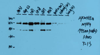
Species tested
Human,Mouse,Rat
Application tested
WB
Organization tested
A431,HepG2,U87,HT-29,293T,A549,Brain(M),Kindey(M),Lung(R)
Barcode encoding
Brief protocol
1. Block with 3% skim milk for 1 hour at room temperature.
2. Incubate overnight with Abgent primary antibody 1:1000 in 3% skim milk at 4℃
3. Wash 5*5 min with TBST.
4. Incubate with HRP-conjugated secondary antibody 1:5000 in 3% skim milk for 1 hour at room temperature.
5. Wash 5*5 min with TBST.
6. Incubate with ECL substrates and expose
|
|


