


 癌症的基本特征包括细胞增殖、血管生成、迁移、凋亡逃避机制和细胞永生等。找到癌症发生过程中这些通路的关键标记物和对应的抗体用于检测至关重要。
 为您推荐一个泛素化位点预测神器——泛素化分析工具,可以为您的蛋白的泛素化位点作出预测和评分。
 细胞自噬受体图形绘图工具为你的蛋白的细胞受体结合位点作出预测和评分,识别结合到自噬通路中的蛋白是非常重要的,便于让我们理解自噬在正常生理、病理过程中的作用,如发育、细胞分化、神经退化性疾病、压力条件下、感染和癌症。
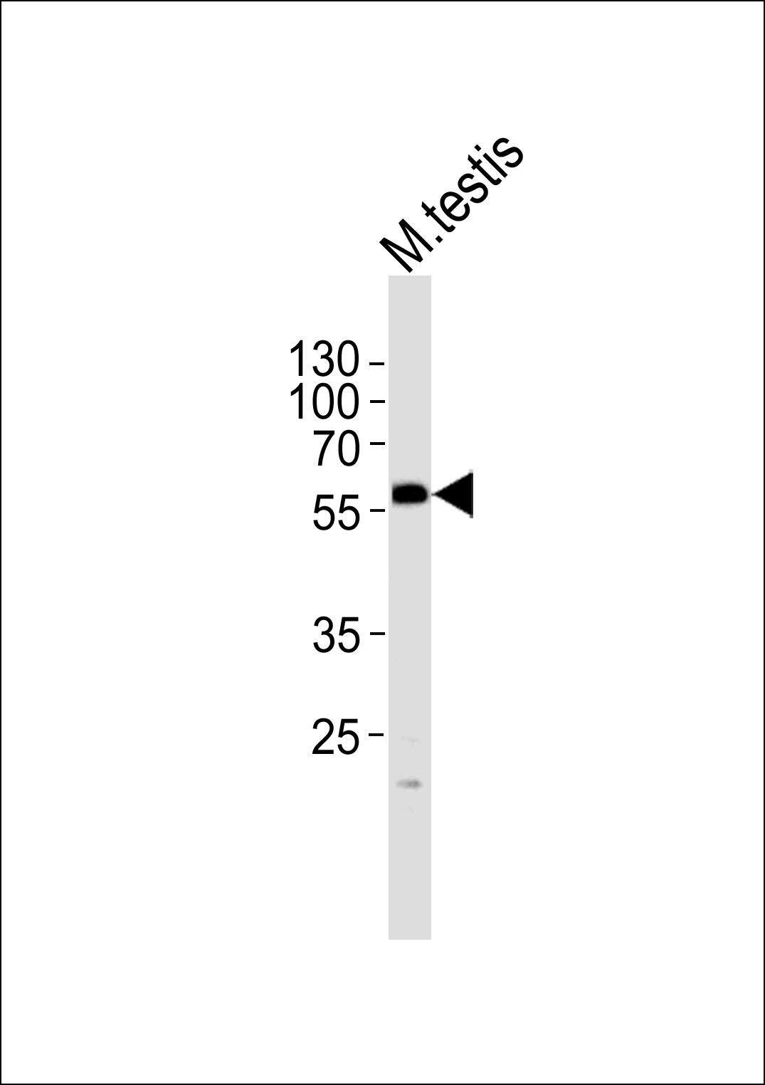
PHF1 Antibody (N-term)
Purified Rabbit Polyclonal Antibody (Pab)
- 产品详情
- 实验流程
- 背景知识
Application
| WB, E |
|---|---|
| Primary Accession | O43189 |
| Reactivity | Human, Rat, Mouse |
| Host | Rabbit |
| Clonality | Polyclonal |
| Isotype | Rabbit IgG |
| Calculated MW | 62106 Da |
| Gene ID | 5252 |
|---|---|
| Other Names | PHD finger protein 1, Protein PHF1, hPHF1, Polycomb-like protein 1, hPCl1, PHF1, PCL1 |
| Target/Specificity | This PHF1 antibody is generated from a rabbit immunized with a KLH conjugated synthetic peptide between 164-197 amino acids from the N-terminal region of human PHF1. |
| Dilution | WB~~1:1000 E~~Use at an assay dependent concentration. |
| Format | Purified polyclonal antibody supplied in PBS with 0.09% (W/V) sodium azide. This antibody is purified through a protein A column, followed by peptide affinity purification. |
| Storage | Maintain refrigerated at 2-8°C for up to 2 weeks. For long term storage store at -20°C in small aliquots to prevent freeze-thaw cycles. |
| Precautions | PHF1 Antibody (N-term) is for research use only and not for use in diagnostic or therapeutic procedures. |
| Name | PHF1 |
|---|---|
| Synonyms | PCL1 |
| Function | Polycomb group (PcG) that specifically binds histone H3 trimethylated at 'Lys-36' (H3K36me3) and recruits the PRC2 complex. Involved in DNA damage response and is recruited at double-strand breaks (DSBs). Acts by binding to H3K36me3, a mark for transcriptional activation, and recruiting the PRC2 complex: it is however unclear whether recruitment of the PRC2 complex to H3K36me3 leads to enhance or inhibit H3K27me3 methylation mediated by the PRC2 complex. According to some reports, PRC2 recruitment by PHF1 promotes H3K27me3 and subsequent gene silencing by inducing spreading of PRC2 and H3K27me3 into H3K36me3 loci (PubMed:18285464, PubMed:23273982). According to another report, PHF1 recruits the PRC2 complex at double-strand breaks (DSBs) and inhibits the activity of PRC2 (PubMed:23142980). Regulates p53/TP53 stability and prolonges its turnover: may act by specifically binding to a methylated from of p53/TP53. |
| Cellular Location | Nucleus. Cytoplasm, cytoskeleton, microtubule organizing center, centrosome. Note=Localizes specifically to the promoters of numerous target genes. Localizes to double-strand breaks (DSBs) sites following DNA damage. Co-localizes with NEK6 in the centrosome |
| Tissue Location | Highest levels in heart, skeletal muscle, and pancreas, lower levels in brain, placenta, lung, liver and kidney |
For Research Use Only. Not For Use In Diagnostic Procedures.
Provided below are standard protocols that you may find useful for product applications.
BACKGROUND
Polycomb group (PcG) that specifically binds histone H3 trimethylated at 'Lys-36' (H3K36me3) and recruits the PRC2 complex. Involved in DNA damage response and is recruited at double-strand breaks (DSBs). Acts by binding to H3K36me3, a mark for transcriptional activation, and recruiting the PRC2 complex: it is however unclear whether recruitment of the PRC2 complex to H3K36me3 leads to enhance or inhibit H3K27me3 methylation mediated by the PRC2 complex. According to some reports, PRC2 recruitment by PHF1 promotes H3K27me3 and subsequent gene silencing by inducing spreading of PRC2 and H3K27me3 into H3K36me3 loci (PubMed:18285464 and PubMed:23273982). According to another report, PHF1 recruits the PRC2 complex at double-strand breaks (DSBs) and inhibits the activity of PRC2 (PubMed:23142980). Regulates p53/TP53 stability and prolonges its turnover: may act by specifically binding to a methylated from of p53/TP53.
REFERENCES
Coulson M.,et al.Genomics 48:381-383(1998).
Wang J.H.,et al.Submitted (MAR-1998) to the EMBL/GenBank/DDBJ databases.
Mungall A.J.,et al.Nature 425:805-811(2003).
Mural R.J.,et al.Submitted (JUL-2005) to the EMBL/GenBank/DDBJ databases.
Micci F.,et al.Cancer Res. 66:107-112(2006).
终于等到您。ABCEPTA(百远生物)抗体产品。
点击下方“我要评价 ”按钮提交您的反馈信息,您的反馈和评价是我们最宝贵的财富之一,
我们将在1-3个工作日内处理您的反馈信息。
如有疑问,联系:0512-88856768 tech-china@abcepta.com.


