


 癌症的基本特征包括细胞增殖、血管生成、迁移、凋亡逃避机制和细胞永生等。找到癌症发生过程中这些通路的关键标记物和对应的抗体用于检测至关重要。
 为您推荐一个泛素化位点预测神器——泛素化分析工具,可以为您的蛋白的泛素化位点作出预测和评分。
 细胞自噬受体图形绘图工具为你的蛋白的细胞受体结合位点作出预测和评分,识别结合到自噬通路中的蛋白是非常重要的,便于让我们理解自噬在正常生理、病理过程中的作用,如发育、细胞分化、神经退化性疾病、压力条件下、感染和癌症。
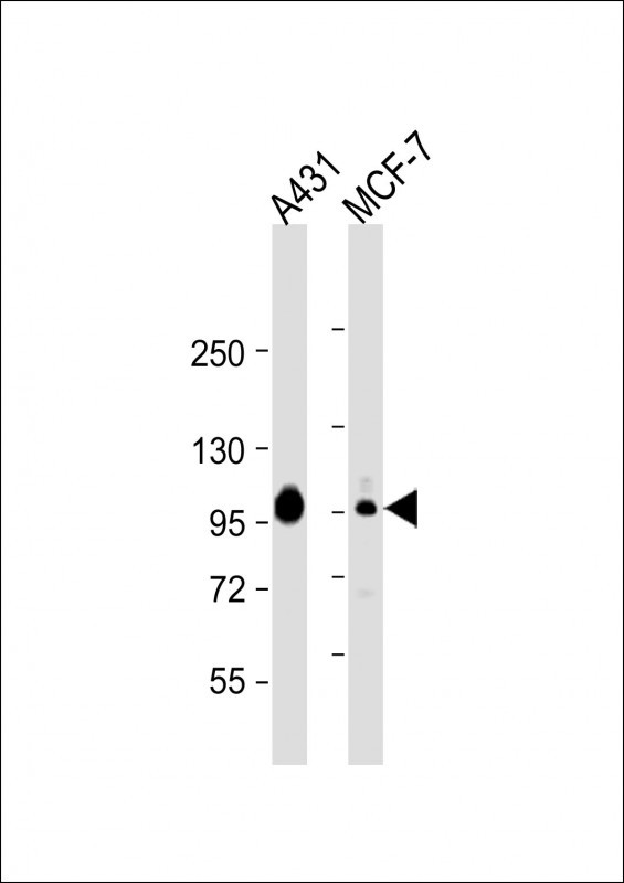
HUMAN-CTNND1_isform 2ABC(Y174) Antibody
Purified Rabbit Polyclonal Antibody (Pab)
- 产品详情
- 实验流程
- 背景知识
Application
| WB, E |
|---|---|
| Primary Accession | O60716 |
| Reactivity | Human, Mouse |
| Host | Rabbit |
| Clonality | Polyclonal |
| Isotype | Rabbit IgG |
| Calculated MW | 108170 Da |
| Gene ID | 1500 |
|---|---|
| Other Names | Catenin delta-1, Cadherin-associated Src substrate, CAS, p120 catenin, p120(ctn), p120(cas), CTNND1, KIAA0384 |
| Target/Specificity | This antibody is generated from a rabbit immunized with a KLH conjugated synthetic peptide between 160-200 amino acids from human. |
| Dilution | WB~~1:2000 E~~Use at an assay dependent concentration. |
| Format | Purified polyclonal antibody supplied in PBS with 0.09% (W/V) sodium azide. This antibody is purified through a protein A column, followed by peptide affinity purification. |
| Storage | Maintain refrigerated at 2-8°C for up to 2 weeks. For long term storage store at -20°C in small aliquots to prevent freeze-thaw cycles. |
| Precautions | HUMAN-CTNND1_isform 2ABC(Y174) Antibody is for research use only and not for use in diagnostic or therapeutic procedures. |
| Name | CTNND1 (HGNC:2515) |
|---|---|
| Synonyms | KIAA0384 |
| Function | Key regulator of cell-cell adhesion that associates with and regulates the cell adhesion properties of both C-, E- and N-cadherins, being critical for their surface stability (PubMed:14610055, PubMed:20371349). Promotes localization and retention of DSG3 at cell- cell junctions, via its interaction with DSG3 (PubMed:18343367). Beside cell-cell adhesion, regulates gene transcription through several transcription factors including ZBTB33/Kaiso2 and GLIS2, and the activity of Rho family GTPases and downstream cytoskeletal dynamics (PubMed:10207085, PubMed:20371349). Implicated both in cell transformation by SRC and in ligand-induced receptor signaling through the EGF, PDGF, CSF-1 and ERBB2 receptors (PubMed:17344476). |
| Cellular Location | Cell junction, adherens junction. Cytoplasm. Nucleus. Cell membrane. Cell junction. Note=Interaction with GLIS2 promotes nuclear translocation (By similarity). Detected at cell-cell contacts (PubMed:15240885, PubMed:17047063). NANOS1 induces its translocation from sites of cell-cell contact to the cytoplasm (PubMed:17047063). CDH1 enhances cell membrane localization (PubMed:15240885). Localizes to cell-cell contacts as keratinocyte differentiation progresses (By similarity) {ECO:0000250|UniProtKB:P30999, ECO:0000269|PubMed:11896187, ECO:0000269|PubMed:15240885, ECO:0000269|PubMed:17047063} [Isoform 2A]: Nucleus [Isoform 4A]: Cytoplasm |
| Tissue Location | Expressed in vascular endothelium. Melanocytes and melanoma cells primarily express the long isoform 1A, whereas keratinocytes express shorter isoforms, especially 3A. The shortest isoform 4A, is detected in normal keratinocytes and melanocytes, and generally lost from cells derived from squamous cell carcinomas or melanomas. The C-terminal alternatively spliced exon B is present in the p120ctn transcripts in the colon, intestine and prostate, but lost in several tumor tissues derived from these organs |
For Research Use Only. Not For Use In Diagnostic Procedures.
Provided below are standard protocols that you may find useful for product applications.
BACKGROUND
Binds to and inhibits the transcriptional repressor ZBTB33, which may lead to activation of target genes of the Wnt signaling pathway (By similarity). Associates with and regulates the cell adhesion properties of both C-, E- and N-cadherins, being critical for their surface stability. Implicated both in cell transformation by SRC and in ligand-induced receptor signaling through the EGF, PDGF, CSF-1 and ERBB2 receptors. Promotes GLIS2 C-terminal cleavage.
REFERENCES
Keirsebilck A.,et al.Genomics 50:129-146(1998).
Nagase T.,et al.DNA Res. 4:141-150(1997).
Ota T.,et al.Nat. Genet. 36:40-45(2004).
Taylor T.D.,et al.Nature 440:497-500(2006).
Kim L.,et al.Mol. Cell. Biol. 15:4553-4561(1995).
终于等到您。ABCEPTA(百远生物)抗体产品。
点击下方“我要评价 ”按钮提交您的反馈信息,您的反馈和评价是我们最宝贵的财富之一,
我们将在1-3个工作日内处理您的反馈信息。
如有疑问,联系:0512-88856768 tech-china@abcepta.com.


