


 癌症的基本特征包括细胞增殖、血管生成、迁移、凋亡逃避机制和细胞永生等。找到癌症发生过程中这些通路的关键标记物和对应的抗体用于检测至关重要。
 为您推荐一个泛素化位点预测神器——泛素化分析工具,可以为您的蛋白的泛素化位点作出预测和评分。
 细胞自噬受体图形绘图工具为你的蛋白的细胞受体结合位点作出预测和评分,识别结合到自噬通路中的蛋白是非常重要的,便于让我们理解自噬在正常生理、病理过程中的作用,如发育、细胞分化、神经退化性疾病、压力条件下、感染和癌症。
(DANRE) rho Antibody (N-Term)
Purified Rabbit Polyclonal Antibody (Pab)
- 产品详情
- 实验流程
- 背景知识
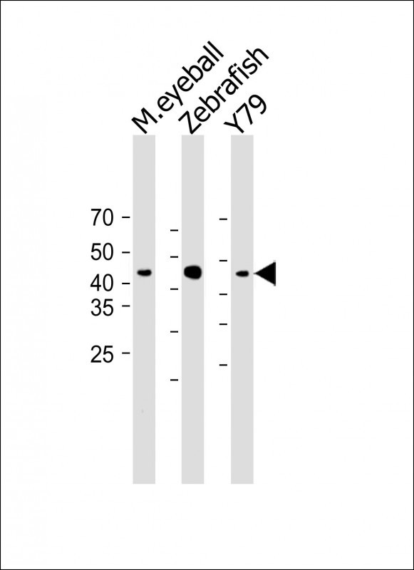
Application
| WB, E |
|---|---|
| Primary Accession | P35359 |
| Reactivity | Zebrafish |
| Host | Rabbit |
| Clonality | polyclonal |
| Isotype | Rabbit IgG |
| Calculated MW | 39706 Da |
| Gene ID | 30295 |
|---|---|
| Other Names | Rhodopsin, rho, zfo2 |
| Target/Specificity | This DANRE rho antibody is generated from a rabbit immunized with a KLH conjugated synthetic peptide between 62-93 amino acids from the human region of DANRE rho. |
| Dilution | WB~~1:1000 E~~Use at an assay dependent concentration. |
| Format | Purified polyclonal antibody supplied in PBS with 0.05% (V/V) Proclin 300. This antibody is purified through a protein A column, followed by peptide affinity purification. |
| Storage | Maintain refrigerated at 2-8°C for up to 2 weeks. For long term storage store at -20°C in small aliquots to prevent freeze-thaw cycles. |
| Precautions | (DANRE) rho Antibody (N-Term) is for research use only and not for use in diagnostic or therapeutic procedures. |
| Name | rho |
|---|---|
| Synonyms | zfo2 |
| Function | Photoreceptor required for image-forming vision at low light intensity. While most salt water fish species use retinal as chromophore, most freshwater fish use 3-dehydroretinal, or a mixture of retinal and 3-dehydroretinal (By similarity). Light-induced isomerization of 11-cis to all-trans retinal triggers a conformational change that activates signaling via G-proteins. Subsequent receptor phosphorylation mediates displacement of the bound G-protein alpha subunit by arrestin and terminates signaling (By similarity). |
| Cellular Location | Membrane {ECO:0000250|UniProtKB:P08100}; Multi- pass membrane protein {ECO:0000250|UniProtKB:P08100}. Cell projection, cilium, photoreceptor outer segment Note=Synthesized in the inner segment (IS) of rod photoreceptor cells before vectorial transport to disk membranes in the rod outer segment (OS) photosensory cilia. {ECO:0000250|UniProtKB:P08100} |
| Tissue Location | Retinal rod photoreceptor cells, predominantly in the outer segments (at protein level) (PubMed:10349976). Retinal rod photoreceptor cells (PubMed:8327475, PubMed:8603882) |
For Research Use Only. Not For Use In Diagnostic Procedures.
Provided below are standard protocols that you may find useful for product applications.
BACKGROUND
Visual pigments such as rhodopsin and porphyropsin are light-absorbing molecules that mediate vision. Rhodopsin consists of an apoprotein, opsin, covalently linked to 11-cis-retinal. This receptor is coupled to the activation of phospholipase C. Porphyropsin consists of opsin covalently linked to 11-cis 3,4- didehydroretinal.
REFERENCES
Robinson J.,et al.Proc. Natl. Acad. Sci. U.S.A. 90:6009-6012(1993).
Robinson J.,et al.Vis. Neurosci. 12:895-906(1995).
Schmitt E.A.,et al.Vis. Neurosci. 16:601-605(1999).
Vihtelic T.S.,et al.Vis. Neurosci. 16:571-585(1999).
Kennedy B.N.,et al.J. Biol. Chem. 276:14037-14043(2001).
终于等到您。ABCEPTA(百远生物)抗体产品。
点击下方“我要评价 ”按钮提交您的反馈信息,您的反馈和评价是我们最宝贵的财富之一,
我们将在1-3个工作日内处理您的反馈信息。
如有疑问,联系:0512-88856768 tech-china@abcepta.com.


