


 癌症的基本特征包括细胞增殖、血管生成、迁移、凋亡逃避机制和细胞永生等。找到癌症发生过程中这些通路的关键标记物和对应的抗体用于检测至关重要。
 为您推荐一个泛素化位点预测神器——泛素化分析工具,可以为您的蛋白的泛素化位点作出预测和评分。
 细胞自噬受体图形绘图工具为你的蛋白的细胞受体结合位点作出预测和评分,识别结合到自噬通路中的蛋白是非常重要的,便于让我们理解自噬在正常生理、病理过程中的作用,如发育、细胞分化、神经退化性疾病、压力条件下、感染和癌症。
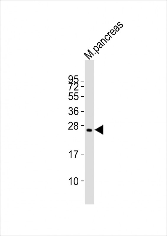
(Mouse) Plet1 Antibody (Center)
Purified Rabbit Polyclonal Antibody (Pab)
- 产品详情
- 实验流程
- 背景知识
Application
| WB, E |
|---|---|
| Primary Accession | Q8VEN2 |
| Reactivity | Mouse |
| Host | Rabbit |
| Clonality | polyclonal |
| Isotype | Rabbit IgG |
| Calculated MW | 25006 Da |
| Gene ID | 76509 |
|---|---|
| Other Names | Placenta-expressed transcript 1 protein, Antigen mAgK114, Plet1 |
| Target/Specificity | This Mouse Plet1 antibody is generated from a rabbit immunized with a KLH conjugated synthetic peptide between 82-115 amino acids from the Central region of Mouse Plet1. |
| Dilution | WB~~1:2000 E~~Use at an assay dependent concentration. |
| Format | Purified polyclonal antibody supplied in PBS with 0.05% (V/V) Proclin 300. This antibody is prepared by Saturated Ammonium Sulfate (SAS) precipitation followed by dialysis against PBS. |
| Storage | Maintain refrigerated at 2-8°C for up to 2 weeks. For long term storage store at -20°C in small aliquots to prevent freeze-thaw cycles. |
| Precautions | (Mouse) Plet1 Antibody (Center) is for research use only and not for use in diagnostic or therapeutic procedures. |
| Name | Plet1 |
|---|---|
| Function | Modulates leading keratinocyte migration and cellular adhesion to matrix proteins during a wound-healing response and promotes wound repair. May play a role during trichilemmal differentiation of the hair follicle. |
| Cellular Location | Apical cell membrane; Lipid-anchor, GPI-anchor. Note=Localized at the apical membrane of the most differentiated keratinocytes of the outer root sheath (ORS), clustered mainly in planar regions of the plasma membrane at the base of microvilli |
| Tissue Location | Present in hair follicle cells and sebaceous gland of skin, ciliated epithelial cells of trachea and bronchial tube, striated portion of submandibular gland, distal convoluted tubule cells of kidney, ciliated epithelial cells of oviduct, medulla of adrenal gland and anterior lobe of pituitary gland. Expressed in keratinocytes of the hair follicle at the trichilemmal zone corresponding to the terminally differentiated outermost suprabasal outer root sheath (ORS), including that of the sebaceous gland duct (SGD) and the directly adjacent upper distal end of the companion layer (CL). Expression is similar in all hair follicle growth stages. Also detected during both the early and late anagen phases above the bulge of stem cells Expressed at the leading edge of the epidermal wound. Not expressed in the interfollicular epidermis (IFE), inner root sheath (IRS) and hair fiber. Highly expressed in placenta. Detected in mammary and prostate epithelia and in the pancreas (at protein level) |
For Research Use Only. Not For Use In Diagnostic Procedures.
Provided below are standard protocols that you may find useful for product applications.
BACKGROUND
Modulates leading keratinocyte migration and cellular adhesion to matrix proteins during a wound-healing response and promotes wound repair. May play a role during trichilemmal differentiation of the hair follicle.
REFERENCES
Zhao S.-H.,et al.Genomics 84:114-125(2004).
Carninci P.,et al.Science 309:1559-1563(2005).
Takeuchi M.,et al.Zool. Sci. 22:995-1001(2005).
Tatefuji T.,et al.Biol. Pharm. Bull. 29:896-902(2006).
Frankenberg S.,et al.BMC Dev. Biol. 7:8-8(2007).
终于等到您。ABCEPTA(百远生物)抗体产品。
点击下方“我要评价 ”按钮提交您的反馈信息,您的反馈和评价是我们最宝贵的财富之一,
我们将在1-3个工作日内处理您的反馈信息。
如有疑问,联系:0512-88856768 tech-china@abcepta.com.


