


 癌症的基本特征包括细胞增殖、血管生成、迁移、凋亡逃避机制和细胞永生等。找到癌症发生过程中这些通路的关键标记物和对应的抗体用于检测至关重要。
 为您推荐一个泛素化位点预测神器——泛素化分析工具,可以为您的蛋白的泛素化位点作出预测和评分。
 细胞自噬受体图形绘图工具为你的蛋白的细胞受体结合位点作出预测和评分,识别结合到自噬通路中的蛋白是非常重要的,便于让我们理解自噬在正常生理、病理过程中的作用,如发育、细胞分化、神经退化性疾病、压力条件下、感染和癌症。
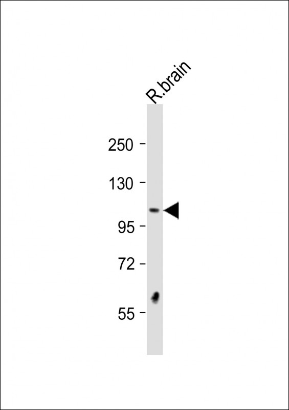
DSTYK Antibody (N-Term)
Purified Rabbit Polyclonal Antibody (Pab)
- 产品详情
- 实验流程
- 背景知识
Application
| WB, E |
|---|---|
| Primary Accession | Q6XUX3 |
| Reactivity | Human, Rat |
| Host | Rabbit |
| Clonality | polyclonal |
| Isotype | Rabbit IgG |
| Calculated MW | 105206 Da |
| Gene ID | 25778 |
|---|---|
| Other Names | Dual serine/threonine and tyrosine protein kinase, Dusty protein kinase, Dusty PK, RIP-homologous kinase, Receptor-interacting serine/threonine-protein kinase 5, Sugen kinase 496, SgK496, DSTYK, KIAA0472, RIP5, RIPK5, SGK496 |
| Target/Specificity | This DSTYK antibody is generated from a rabbit immunized with a KLH conjugated synthetic peptide between 256-290 amino acids from human DSTYK. |
| Dilution | WB~~1:2000 E~~Use at an assay dependent concentration. |
| Format | Purified polyclonal antibody supplied in PBS with 0.09% (W/V) sodium azide. This antibody is purified through a protein A column, followed by peptide affinity purification. |
| Storage | Maintain refrigerated at 2-8°C for up to 2 weeks. For long term storage store at -20°C in small aliquots to prevent freeze-thaw cycles. |
| Precautions | DSTYK Antibody (N-Term) is for research use only and not for use in diagnostic or therapeutic procedures. |
| Name | DSTYK |
|---|---|
| Synonyms | KIAA0472, RIP5, RIPK5, SGK496 |
| Function | Acts as a positive regulator of ERK phosphorylation downstream of fibroblast growth factor-receptor activation (PubMed:23862974, PubMed:28157540). Involved in the regulation of both caspase-dependent apoptosis and caspase-independent cell death (PubMed:15178406). In the skin, it plays a predominant role in suppressing caspase-dependent apoptosis in response to UV stress in a range of dermal cell types (PubMed:28157540). |
| Cellular Location | Cytoplasm. Cell membrane {ECO:0000250|UniProtKB:Q6XUX1}. Apical cell membrane. Basolateral cell membrane. Cell junction {ECO:0000250|UniProtKB:Q6XUX1}. Note=Detected at apical cell-cell junctions. Colocalized with FGF receptors to the cell membrane (By similarity). Detected in basolateral and apical membranes of all tubular epithelia. {ECO:0000250|UniProtKB:Q6XUX1, ECO:0000269|PubMed:23862974} |
| Tissue Location | Predominantly expressed in skeletal muscle and testis. Expressed in basolateral and apical membranes of all tubular epithelia. Expressed in thin ascending limb of the loop of Henle and the distal convoluted tubule. Expressed in all layers of transitional ureteric epithelium and in the ureteric smooth-muscle cells. Weakly expressed in heart, brain, placenta, kidney, pancreas, spleen, thymus, prostate, uterus, small intestine, white blood cells, stomach, spinal cord and adrenal gland. Is widely distributed in the CNS. Also detected in several tumor cell lines. Expressed in the skin (PubMed:28157540) |
For Research Use Only. Not For Use In Diagnostic Procedures.
Provided below are standard protocols that you may find useful for product applications.
BACKGROUND
Acts as a positive regulator of ERK phosphorylation downstream of fibroblast growth factor-receptor activation. May induce both caspase-dependent apoptosis and caspase-independent cell death.
REFERENCES
Peng J.,et al.Biochim. Biophys. Acta 1759:562-572(2006).
Zhao Z.,et al.Submitted (MAY-1998) to the EMBL/GenBank/DDBJ databases.
Gregory S.G.,et al.Nature 441:315-321(2006).
Seki N.,et al.DNA Res. 4:345-349(1997).
Zha J.,et al.Biochem. Biophys. Res. Commun. 319:298-303(2004).
终于等到您。ABCEPTA(百远生物)抗体产品。
点击下方“我要评价 ”按钮提交您的反馈信息,您的反馈和评价是我们最宝贵的财富之一,
我们将在1-3个工作日内处理您的反馈信息。
如有疑问,联系:0512-88856768 tech-china@abcepta.com.


