


 癌症的基本特征包括细胞增殖、血管生成、迁移、凋亡逃避机制和细胞永生等。找到癌症发生过程中这些通路的关键标记物和对应的抗体用于检测至关重要。
 为您推荐一个泛素化位点预测神器——泛素化分析工具,可以为您的蛋白的泛素化位点作出预测和评分。
 细胞自噬受体图形绘图工具为你的蛋白的细胞受体结合位点作出预测和评分,识别结合到自噬通路中的蛋白是非常重要的,便于让我们理解自噬在正常生理、病理过程中的作用,如发育、细胞分化、神经退化性疾病、压力条件下、感染和癌症。
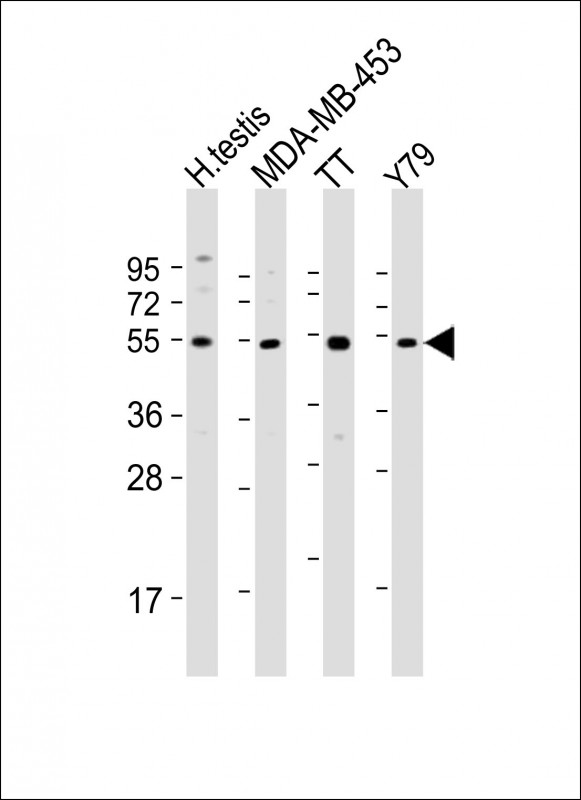
WDR51B Antibody (C-term)
Purified Rabbit Polyclonal Antibody (Pab)
- 产品详情
- 实验流程
- 背景知识
Application
| WB, E |
|---|---|
| Primary Accession | Q8TC44 |
| Reactivity | Human |
| Host | Rabbit |
| Clonality | polyclonal |
| Isotype | Rabbit IgG |
| Calculated MW | 53668 Da |
| Gene ID | 282809 |
|---|---|
| Other Names | POC1 centriolar protein homolog B, Pix1, Proteome of centriole protein 1B, WD repeat-containing protein 51B, POC1B, WDR51B |
| Target/Specificity | This WDR51B antibody is generated from a rabbit immunized with a KLH conjugated synthetic peptide between 386-417 amino acids from the C-terminal region of human WDR51B. |
| Dilution | WB~~1:2000 E~~Use at an assay dependent concentration. |
| Format | Purified polyclonal antibody supplied in PBS with 0.09% (W/V) sodium azide. This antibody is purified through a protein A column, followed by peptide affinity purification. |
| Storage | Maintain refrigerated at 2-8°C for up to 2 weeks. For long term storage store at -20°C in small aliquots to prevent freeze-thaw cycles. |
| Precautions | WDR51B Antibody (C-term) is for research use only and not for use in diagnostic or therapeutic procedures. |
| Name | POC1B (HGNC:30836) |
|---|---|
| Synonyms | WDR51B |
| Function | Plays an important role in centriole assembly and/or stability and ciliogenesis (PubMed:20008567, PubMed:32060285). Involved in early steps of centriole duplication, as well as in the later steps of centriole length control (PubMed:19109428). Acts in concert with POC1A to ensure centriole integrity and proper mitotic spindle formation (PubMed:32060285). Required for primary cilia formation, ciliary length and also cell proliferation (PubMed:23015594). Required for retinal integrity (PubMed:25044745). Acts as a positive regulator of centriole elongation (PubMed:37934472). |
| Cellular Location | Cytoplasm, cytoskeleton, microtubule organizing center, centrosome. Cytoplasm, cytoskeleton, microtubule organizing center, centrosome, centriole. Cytoplasm, cytoskeleton, cilium basal body Cytoplasm, cytoskeleton, spindle pole. Note=Component of both mother and daughter centrioles (PubMed:32060285). Localizes to the basal body and centriole adjacent to the connecting cilium of photoreceptors and in synapses of the outer plexiform layer. Localizes to the inner scaffold in the central region of centrioles {ECO:0000250|UniProtKB:Q8BHD1, ECO:0000269|PubMed:32060285, ECO:0000269|PubMed:37934472} |
| Tissue Location | Expressed in the retina. |
For Research Use Only. Not For Use In Diagnostic Procedures.
Provided below are standard protocols that you may find useful for product applications.
BACKGROUND
Plays an important role in centriole assembly and/or stability and ciliogenesis (PubMed:20008567). Involved in early steps of centriole duplication, as well as in the later steps of centriole length control (PubMed:19109428). Acts in concert with POC1A to ensure centriole integrity and proper mitotic spindle formation. Required for primary cilia formation, ciliary length and also cell proliferation (PubMed:23015594). Required for retinal integrity (PubMed:25044745).
REFERENCES
Ota T.,et al.Nat. Genet. 36:40-45(2004).
Scherer S.E.,et al.Nature 440:346-351(2006).
Mural R.J.,et al.Submitted (JUL-2005) to the EMBL/GenBank/DDBJ databases.
Hames R.S.,et al.Exp. Cell Res. 314:574-589(2008).
Dephoure N.,et al.Proc. Natl. Acad. Sci. U.S.A. 105:10762-10767(2008).
终于等到您。ABCEPTA(百远生物)抗体产品。
点击下方“我要评价 ”按钮提交您的反馈信息,您的反馈和评价是我们最宝贵的财富之一,
我们将在1-3个工作日内处理您的反馈信息。
如有疑问,联系:0512-88856768 tech-china@abcepta.com.


