


 癌症的基本特征包括细胞增殖、血管生成、迁移、凋亡逃避机制和细胞永生等。找到癌症发生过程中这些通路的关键标记物和对应的抗体用于检测至关重要。
 为您推荐一个泛素化位点预测神器——泛素化分析工具,可以为您的蛋白的泛素化位点作出预测和评分。
 细胞自噬受体图形绘图工具为你的蛋白的细胞受体结合位点作出预测和评分,识别结合到自噬通路中的蛋白是非常重要的,便于让我们理解自噬在正常生理、病理过程中的作用,如发育、细胞分化、神经退化性疾病、压力条件下、感染和癌症。
SYVN1 (HRD1) Antibody (N-term)
Purified Rabbit Polyclonal Antibody (Pab)
- 产品详情
- 文献引用 : 3
- 实验流程
- 背景知识
Application
| WB, E |
|---|---|
| Primary Accession | Q86TM6 |
| Other Accession | Q5XHH7, Q6NRL6, Q9DBY1, Q8N6E8 |
| Reactivity | Human, Mouse |
| Predicted | Mouse, Xenopus |
| Host | Rabbit |
| Clonality | Polyclonal |
| Isotype | Rabbit IgG |
| Calculated MW | 67685 Da |
| Antigen Region | 58-88 aa |
| Gene ID | 84447 |
|---|---|
| Other Names | E3 ubiquitin-protein ligase synoviolin, 632-, Synovial apoptosis inhibitor 1, SYVN1, HRD1, KIAA1810 |
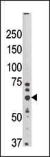
| Target/Specificity | This SYVN1 (HRD1) antibody is generated from rabbits immunized with a KLH conjugated synthetic peptide between 58-88 amino acids from the N-terminal region of human SYVN1 (HRD1). |
| Dilution | WB~~1:1000 E~~Use at an assay dependent concentration. |
| Format | Purified polyclonal antibody supplied in PBS with 0.09% (W/V) sodium azide. This antibody is prepared by Saturated Ammonium Sulfate (SAS) precipitation followed by dialysis against PBS. |
| Storage | Maintain refrigerated at 2-8°C for up to 2 weeks. For long term storage store at -20°C in small aliquots to prevent freeze-thaw cycles. |
| Precautions | SYVN1 (HRD1) Antibody (N-term) is for research use only and not for use in diagnostic or therapeutic procedures. |
| Name | SYVN1 {ECO:0000303|PubMed:15489334} |
|---|---|
| Function | E3 ubiquitin-protein ligase which accepts ubiquitin specifically from endoplasmic reticulum-associated UBC7 E2 ligase and transfers it to substrates, promoting their degradation (PubMed:12459480, PubMed:12646171, PubMed:12975321, PubMed:14593114, PubMed:16289116, PubMed:16847254, PubMed:17059562, PubMed:17141218, PubMed:17170702, PubMed:22607976, PubMed:27827840, PubMed:26471130, PubMed:28827405). Component of the endoplasmic reticulum quality control (ERQC) system also called ER-associated degradation (ERAD) involved in ubiquitin-dependent degradation of misfolded endoplasmic reticulum proteins (PubMed:12459480, PubMed:12646171, PubMed:12975321, PubMed:14593114, PubMed:16289116, PubMed:16847254, PubMed:17059562, PubMed:17141218, PubMed:17170702, PubMed:22607976, PubMed:26471130, PubMed:28842558). Also promotes the degradation of normal but naturally short-lived proteins such as SGK. Protects cells from ER stress-induced apoptosis. Protects neurons from apoptosis induced by polyglutamine- expanded huntingtin (HTT) or unfolded GPR37 by promoting their degradation (PubMed:17141218). Sequesters p53/TP53 in the cytoplasm and promotes its degradation, thereby negatively regulating its biological function in transcription, cell cycle regulation and apoptosis (PubMed:17170702). Mediates the ubiquitination and subsequent degradation of cytoplasmic NFE2L1 (By similarity). During the early stage of B cell development, required for degradation of the pre-B cell receptor (pre-BCR) complex, hence supporting further differentiation into mature B cells (By similarity). |
| Cellular Location | Endoplasmic reticulum membrane; Multi-pass membrane protein |
| Tissue Location | Ubiquitously expressed, with highest levels in liver and kidney (at protein level). Up-regulated in synovial tissues from patients with rheumatoid arthritis (at protein level) |
For Research Use Only. Not For Use In Diagnostic Procedures.

Provided below are standard protocols that you may find useful for product applications.
BACKGROUND
HRD1 is a ubiquitin ligase whose expression is induced by the unfolded protein response (UPR) following endoplasmic reticulum stress. Expression of HRD1 protects cells from apoptosis by inducing degradation of abnormally processed proteins that accumulate in the endoplasmic reticulum. HRD1 is expressed in many tissues, strongly expressed in brain, pancreas, liver, kidney and skeletal muscle. Synoviolin/Hrd1 (expressed in rheumatoid synovium) is reported to be a novel causative factor for arthropathy by triggering synovial cell outgrowth through its antiapoptotic effects. HRD1 contains one ring-type zinc finger.
REFERENCES
Kikkert, M., et al., J. Biol. Chem. 279(5):3525-3534 (2004). Amano, T., et al., Genes Dev. 17(19):2436-2449 (2003). Nadav, E., et al., Biochem. Biophys. Res. Commun. 303(1):91-97 (2003). Kaneko, M., et al., FEBS Lett. 532 (1-2), 147-152 (2002) (): ().
终于等到您。ABCEPTA(百远生物)抗体产品。
点击下方“我要评价 ”按钮提交您的反馈信息,您的反馈和评价是我们最宝贵的财富之一,
我们将在1-3个工作日内处理您的反馈信息。
如有疑问,联系:0512-88856768 tech-china@abcepta.com.


