


 癌症的基本特征包括细胞增殖、血管生成、迁移、凋亡逃避机制和细胞永生等。找到癌症发生过程中这些通路的关键标记物和对应的抗体用于检测至关重要。
 为您推荐一个泛素化位点预测神器——泛素化分析工具,可以为您的蛋白的泛素化位点作出预测和评分。
 细胞自噬受体图形绘图工具为你的蛋白的细胞受体结合位点作出预测和评分,识别结合到自噬通路中的蛋白是非常重要的,便于让我们理解自噬在正常生理、病理过程中的作用,如发育、细胞分化、神经退化性疾病、压力条件下、感染和癌症。
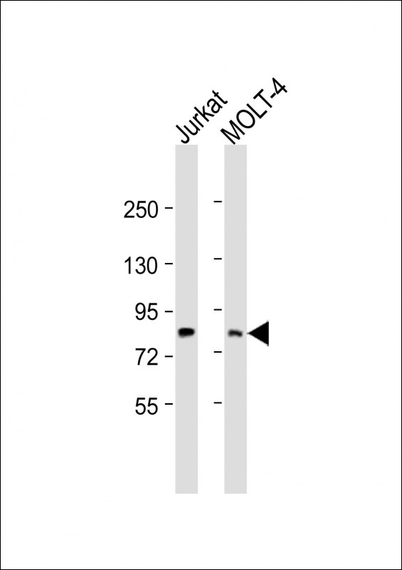
LCP2 Antibody (Center)
Purified Rabbit Polyclonal Antibody (Pab)
- 产品详情
- 实验流程
- 背景知识
Application
| WB, E |
|---|---|
| Primary Accession | Q13094 |
| Other Accession | Q60787 |
| Reactivity | Human, Mouse |
| Predicted | Mouse |
| Host | Rabbit |
| Clonality | polyclonal |
| Isotype | Rabbit IgG |
| Calculated MW | 60188 Da |
| Gene ID | 3937 |
|---|---|
| Other Names | Lymphocyte cytosolic protein 2, SH2 domain-containing leukocyte protein of 76 kDa, SLP-76 tyrosine phosphoprotein, SLP76, LCP2 |
| Target/Specificity | This LCP2 antibody is generated from a rabbit immunized with a KLH conjugated synthetic peptide between 303-337 amino acids from the Central region of human LCP2. |
| Dilution | WB~~1:2000 E~~Use at an assay dependent concentration. |
| Format | Purified polyclonal antibody supplied in PBS with 0.09% (W/V) sodium azide. This antibody is purified through a protein A column, followed by peptide affinity purification. |
| Storage | Maintain refrigerated at 2-8°C for up to 2 weeks. For long term storage store at -20°C in small aliquots to prevent freeze-thaw cycles. |
| Precautions | LCP2 Antibody (Center) is for research use only and not for use in diagnostic or therapeutic procedures. |
| Name | LCP2 |
|---|---|
| Function | Adapter protein primarily involved in signaling pathways within T-cells, as well as other immune cells such as platelets, mast cells, and natural killer (NK) cells (PubMed:11313406, PubMed:33159816). Plays a crucial role for transducing signal from the T-cell receptor (TCR) after antigen recognition leading to T-cell activation. Mechanistically, once phosphorylated by the kinase ZAP70, mediates interactions with the guanine-nucleotide exchange factor VAV1, the adapter protein NCK and the kinase ITK (PubMed:8673706, PubMed:8702662). In turn, stimulates the activation of PKC-theta/PRKCQ and NF-kappa-B transcriptional activity in response to CD3 and CD28 costimulation (PubMed:11313406). Also plays an essential role in AGER- induced signaling pathways including p38 MAPK and ERK1/2 activation leading to cytokine release and pro-inflammatory responses (PubMed:33436632). |
| Cellular Location | Cytoplasm. |
| Tissue Location | Highly expressed in spleen, thymus and peripheral blood leukocytes. Highly expressed also in T-cell and monocytic cell lines, expressed at lower level in B-cell lines. Not detected in fibroblast or neuroblastoma cell lines |
Research Areas
For Research Use Only. Not For Use In Diagnostic Procedures.
Application Protocols
Provided below are standard protocols that you may find useful for product applications.
BACKGROUND
Involved in T-cell antigen receptor mediated signaling.
REFERENCES
Jackman J.K.,et al.J. Biol. Chem. 270:7029-7032(1995).
Kalnine N.,et al.Submitted (MAY-2003) to the EMBL/GenBank/DDBJ databases.
Ota T.,et al.Nat. Genet. 36:40-45(2004).
Mural R.J.,et al.Submitted (SEP-2005) to the EMBL/GenBank/DDBJ databases.
Moog-Lutz C.,et al.J. Biol. Chem. 276:22375-22381(2001).
终于等到您。ABCEPTA(百远生物)抗体产品。
点击下方“我要评价 ”按钮提交您的反馈信息,您的反馈和评价是我们最宝贵的财富之一,
我们将在1-3个工作日内处理您的反馈信息。
如有疑问,联系:0512-88856768 tech-china@abcepta.com.
¥ 1,250.00
Cat# AP22146c


