


 癌症的基本特征包括细胞增殖、血管生成、迁移、凋亡逃避机制和细胞永生等。找到癌症发生过程中这些通路的关键标记物和对应的抗体用于检测至关重要。
 为您推荐一个泛素化位点预测神器——泛素化分析工具,可以为您的蛋白的泛素化位点作出预测和评分。
 细胞自噬受体图形绘图工具为你的蛋白的细胞受体结合位点作出预测和评分,识别结合到自噬通路中的蛋白是非常重要的,便于让我们理解自噬在正常生理、病理过程中的作用,如发育、细胞分化、神经退化性疾病、压力条件下、感染和癌症。
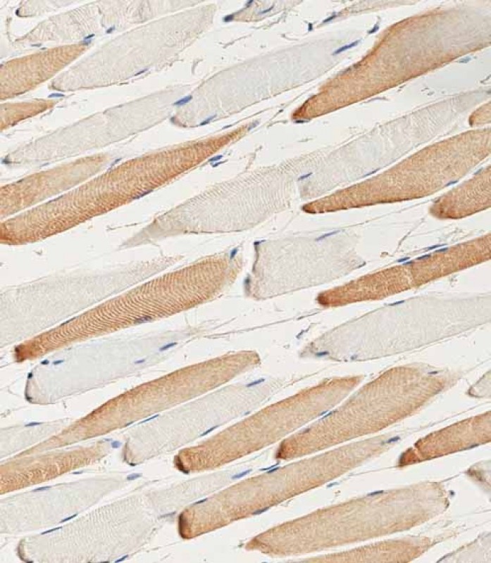
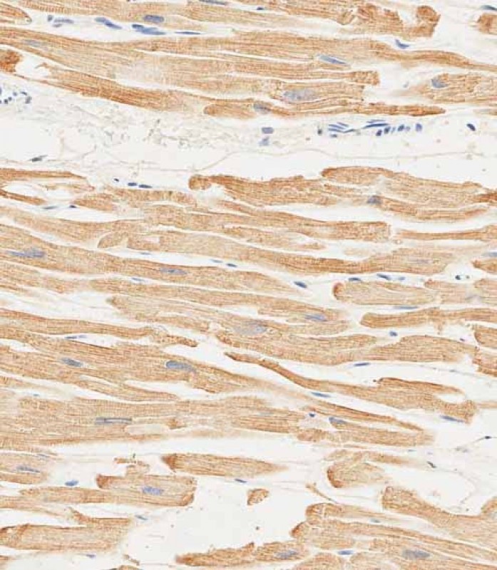
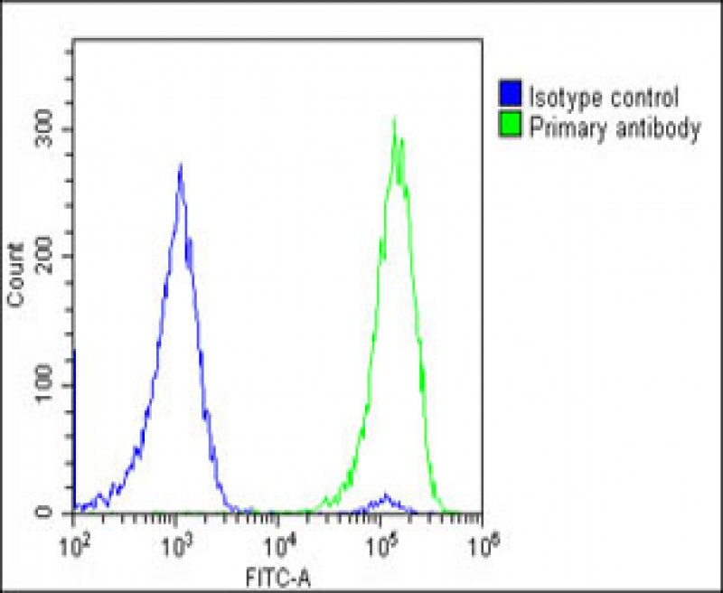
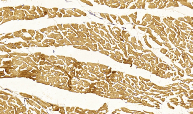
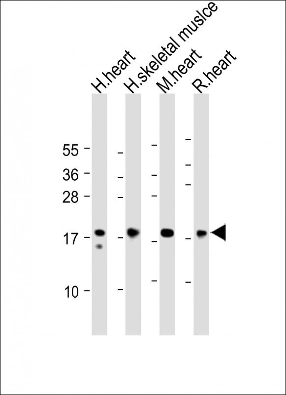
MYL2 Antibody (Center)
Purified Rabbit Polyclonal Antibody (Pab)
- 产品详情
- 实验流程
Application
| WB, FC, IHC-P-Leica, E |
|---|---|
| Primary Accession | P10916 |
| Other Accession | P02610, P51667, Q7M2V4, P08733 |
| Reactivity | Human, Rat, Mouse |
| Predicted | Chicken, Mouse, Rat |
| Host | Rabbit |
| Clonality | polyclonal |
| Isotype | Rabbit IgG |
| Calculated MW | 18789 Da |
| Gene ID | 4633 |
|---|---|
| Other Names | Myosin regulatory light chain 2, ventricular/cardiac muscle isoform, MLC-2, MLC-2v, MYL2 |
| Target/Specificity | This MYL2 antibody is generated from a rabbit immunized with a KLH conjugated synthetic peptide between 42-75 amino acids from the Central region of human MYL2. |
| Dilution | WB~~1:2000 FC~~1:25 IHC-P-Leica~~1:1000 E~~Use at an assay dependent concentration. |
| Format | Purified polyclonal antibody supplied in PBS with 0.09% (W/V) sodium azide. This antibody is purified through a protein A column, followed by peptide affinity purification. |
| Storage | Maintain refrigerated at 2-8°C for up to 2 weeks. For long term storage store at -20°C in small aliquots to prevent freeze-thaw cycles. |
| Precautions | MYL2 Antibody (Center) is for research use only and not for use in diagnostic or therapeutic procedures. |
| Name | MYL2 (HGNC:7583) |
|---|---|
| Function | Contractile protein that plays a role in heart development and function (PubMed:23365102, PubMed:32453731). Following phosphorylation, plays a role in cross-bridge cycling kinetics and cardiac muscle contraction by increasing myosin lever arm stiffness and promoting myosin head diffusion; as a consequence of the increase in maximum contraction force and calcium sensitivity of contraction force. These events altogether slow down myosin kinetics and prolong duty cycle resulting in accumulated myosins being cooperatively recruited to actin binding sites to sustain thin filament activation as a means to fine-tune myofilament calcium sensitivity to force (By similarity). During cardiogenesis plays an early role in cardiac contractility by promoting cardiac myofibril assembly (By similarity). |
| Cellular Location | Cytoplasm, myofibril, sarcomere, A band {ECO:0000250|UniProtKB:P08733} |
| Tissue Location | Highly expressed in type I muscle fibers. |
Research Areas
For Research Use Only. Not For Use In Diagnostic Procedures.
Application Protocols
Provided below are standard protocols that you may find useful for product applications.
REFERENCES
Libera L.D.,et al.Nucleic Acids Res. 17:2360-2360(1989).
Wu Q.L.,et al.Submitted (MAR-1996) to the EMBL/GenBank/DDBJ databases.
Margossian S.S.,et al.Submitted (AUG-1997) to the EMBL/GenBank/DDBJ databases.
Wadgaonkar R.,et al.Cell. Mol. Biol. Res. 39:13-26(1993).
Kovalyov L.I.,et al.Electrophoresis 16:1160-1169(1995).
终于等到您。ABCEPTA(百远生物)抗体产品。
点击下方“我要评价 ”按钮提交您的反馈信息,您的反馈和评价是我们最宝贵的财富之一,
我们将在1-3个工作日内处理您的反馈信息。
如有疑问,联系:0512-88856768 tech-china@abcepta.com.
¥ 699.00
Cat# AP22304c


