


 癌症的基本特征包括细胞增殖、血管生成、迁移、凋亡逃避机制和细胞永生等。找到癌症发生过程中这些通路的关键标记物和对应的抗体用于检测至关重要。
 为您推荐一个泛素化位点预测神器——泛素化分析工具,可以为您的蛋白的泛素化位点作出预测和评分。
 细胞自噬受体图形绘图工具为你的蛋白的细胞受体结合位点作出预测和评分,识别结合到自噬通路中的蛋白是非常重要的,便于让我们理解自噬在正常生理、病理过程中的作用,如发育、细胞分化、神经退化性疾病、压力条件下、感染和癌症。
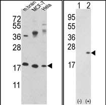
CFL1 Antibody (N-term)
Purified Rabbit Polyclonal Antibody (Pab)
- 产品详情
- 文献引用 : 1
- 实验流程
- 背景知识
Application
| WB, IHC-P, FC, E |
|---|---|
| Primary Accession | P23528 |
| Other Accession | P45592, P10668, P18760, Q4R5C0, Q5E9F7 |
| Reactivity | Human, Mouse |
| Predicted | Bovine, Monkey, Pig, Rat |
| Host | Rabbit |
| Clonality | Polyclonal |
| Isotype | Rabbit IgG |
| Calculated MW | 18502 Da |
| Antigen Region | 4-32 aa |
| Gene ID | 1072 |
|---|---|
| Other Names | Cofilin-1, 18 kDa phosphoprotein, p18, Cofilin, non-muscle isoform, CFL1, CFL |
| Target/Specificity | This CFL1 antibody is generated from rabbits immunized with a KLH conjugated synthetic peptide between 4-32 amino acids from the N-terminal region of human CFL1. |
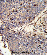
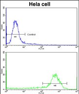
| Dilution | WB~~1:1000 IHC-P~~1:100~500 FC~~1:10~50 E~~Use at an assay dependent concentration. |
| Format | Purified polyclonal antibody supplied in PBS with 0.09% (W/V) sodium azide. This antibody is prepared by Saturated Ammonium Sulfate (SAS) precipitation followed by dialysis against PBS. |
| Storage | Maintain refrigerated at 2-8°C for up to 2 weeks. For long term storage store at -20°C in small aliquots to prevent freeze-thaw cycles. |
| Precautions | CFL1 Antibody (N-term) is for research use only and not for use in diagnostic or therapeutic procedures. |
| Name | CFL1 |
|---|---|
| Synonyms | CFL |
| Function | Binds to F-actin and exhibits pH-sensitive F-actin depolymerizing activity (PubMed:11812157). In conjunction with the subcortical maternal complex (SCMC), plays an essential role for zygotes to progress beyond the first embryonic cell divisions via regulation of actin dynamics (PubMed:15580268). Required for the centralization of the mitotic spindle and symmetric division of zygotes (By similarity). Plays a role in the regulation of cell morphology and cytoskeletal organization in epithelial cells (PubMed:21834987). Required for the up-regulation of atypical chemokine receptor ACKR2 from endosomal compartment to cell membrane, increasing its efficiency in chemokine uptake and degradation (PubMed:23633677). Required for neural tube morphogenesis and neural crest cell migration (By similarity). |
| Cellular Location | Nucleus matrix. Cytoplasm, cytoskeleton. Cell projection, ruffle membrane; Peripheral membrane protein; Cytoplasmic side. Cell projection, lamellipodium membrane; Peripheral membrane protein; Cytoplasmic side. Cell projection, lamellipodium {ECO:0000250|UniProtKB:P18760}. Cell projection, growth cone {ECO:0000250|UniProtKB:P18760}. Cell projection, axon {ECO:0000250|UniProtKB:P18760}. Note=Colocalizes with the actin cytoskeleton in membrane ruffles and lamellipodia. Detected at the cleavage furrow and contractile ring during cytokinesis. Almost completely in nucleus in cells exposed to heat shock or 10% dimethyl sulfoxide |
| Tissue Location | Widely distributed in various tissues. |
Research Areas
For Research Use Only. Not For Use In Diagnostic Procedures.

Application Protocols
Provided below are standard protocols that you may find useful for product applications.
BACKGROUND
Cofilin is a widely distributed intracellular actin-modulating protein that binds and depolymerizes filamentous F-actin and inhibits the polymerization of monomeric G-actin in a pH-dependent manner.
REFERENCES
Fazal,F., et.al., J. Biol. Chem. 284 (31), 21047-21056 (2009)
终于等到您。ABCEPTA(百远生物)抗体产品。
点击下方“我要评价 ”按钮提交您的反馈信息,您的反馈和评价是我们最宝贵的财富之一,
我们将在1-3个工作日内处理您的反馈信息。
如有疑问,联系:0512-88856768 tech-china@abcepta.com.
|
Anonymous
2016-08-24 09:20:44
1
2
3
4
5
|
Species tested
Human,Mouse,Rat
Application tested
WB
Organization tested
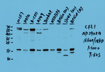
MCF-7,Hela,B-cell,A549,Jurkat,NIH/3T3,Brain(M),Liver(M),Brain(R)
Barcode encoding
Brief protocol
1. Block with 3% skim milk for 1 hour at room temperature.
2. Incubate overnight with Abgent primary antibody 1:1000 in 3% skim milk at 4℃
3. Wash 5*5 min with TBST.
4. Incubate with HRP-conjugated secondary antibody 1:5000 in 3% skim milk for 1 hour at room temperature.
5. Wash 5*5 min with TBST.
6. Incubate with ECL substrates and expose
|
|
¥ 699.00
Cat# AP2905a


