


 癌症的基本特征包括细胞增殖、血管生成、迁移、凋亡逃避机制和细胞永生等。找到癌症发生过程中这些通路的关键标记物和对应的抗体用于检测至关重要。
 为您推荐一个泛素化位点预测神器——泛素化分析工具,可以为您的蛋白的泛素化位点作出预测和评分。
 细胞自噬受体图形绘图工具为你的蛋白的细胞受体结合位点作出预测和评分,识别结合到自噬通路中的蛋白是非常重要的,便于让我们理解自噬在正常生理、病理过程中的作用,如发育、细胞分化、神经退化性疾病、压力条件下、感染和癌症。
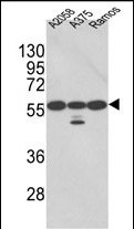
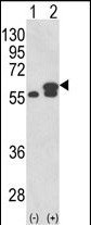
PDIA3 Antibody (Center)
Purified Rabbit Polyclonal Antibody (Pab)
- 产品详情
- 实验流程
- 背景知识
Application
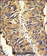
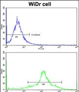
| WB, IHC-P, FC, E |
|---|---|
| Primary Accession | P30101 |
| Other Accession | P38657 |
| Reactivity | Human |
| Predicted | Bovine |
| Host | Rabbit |
| Clonality | Polyclonal |
| Isotype | Rabbit IgG |
| Calculated MW | 56782 Da |
| Antigen Region | 192-220 aa |
| Gene ID | 2923 |
|---|---|
| Other Names | Protein disulfide-isomerase A3, 58 kDa glucose-regulated protein, 58 kDa microsomal protein, p58, Disulfide isomerase ER-60, Endoplasmic reticulum resident protein 57, ER protein 57, ERp57, Endoplasmic reticulum resident protein 60, ER protein 60, ERp60, PDIA3, ERP57, ERP60, GRP58 |
| Target/Specificity | This PDIA3 antibody is generated from rabbits immunized with a KLH conjugated synthetic peptide between 192-220 amino acids from the Central region of human PDIA3. |
| Dilution | WB~~1:1000 IHC-P~~1:100~500 FC~~1:10~50 E~~Use at an assay dependent concentration. |
| Format | Purified polyclonal antibody supplied in PBS with 0.09% (W/V) sodium azide. This antibody is prepared by Saturated Ammonium Sulfate (SAS) precipitation followed by dialysis against PBS. |
| Storage | Maintain refrigerated at 2-8°C for up to 2 weeks. For long term storage store at -20°C in small aliquots to prevent freeze-thaw cycles. |
| Precautions | PDIA3 Antibody (Center) is for research use only and not for use in diagnostic or therapeutic procedures. |
| Name | PDIA3 (HGNC:4606) |
|---|---|
| Synonyms | ERP57, ERP60, GRP58 |
| Function | Protein disulfide isomerase that catalyzes the formation, isomerization, and reduction or oxidation of disulfide bonds in client proteins and functions as a protein folding chaperone (PubMed:11825568, PubMed:16193070, PubMed:27897272, PubMed:36104323, PubMed:7487104). Core component of the major histocompatibility complex class I (MHC I) peptide loading complex where it functions as an essential folding chaperone for TAPBP. Through TAPBP, assists the dynamic assembly of the MHC I complex with high affinity antigens in the endoplasmic reticulum. Therefore, plays a crucial role in the presentation of antigens to cytotoxic T cells in adaptive immunity (PubMed:35948544, PubMed:36104323). |
| Cellular Location | Endoplasmic reticulum. Endoplasmic reticulum lumen {ECO:0000250|UniProtKB:P11598}. Melanosome Note=Identified by mass spectrometry in melanosome fractions from stage I to stage IV (PubMed:12643545). |
| Tissue Location | Detected in the flagellum and head region of spermatozoa (at protein level) (PubMed:20400973). Expressed in liver, stomach and colon (at protein level). Expressed in gastric parietal cells and chief cells (at protein level) (PubMed:24188822) |
For Research Use Only. Not For Use In Diagnostic Procedures.
Provided below are standard protocols that you may find useful for product applications.
BACKGROUND
PDIA3 is the endoplasmic reticulum that interacts with lectin chaperones calreticulin and calnexin to modulate folding of newly synthesized glycoproteins. The protein was once thought to be a phospholipase; however, it has been demonstrated that the protein actually has protein disulfide isomerase activity. It is thought that complexes of lectins and this protein mediate protein folding by promoting formation of disulfide bonds in their glycoprotein substrates.
REFERENCES
Vigneron,N., et.al., Eur. J. Immunol. 39 (9), 2371-2376 (2009)
Xu,D.,et.al., Am. J. Physiol. Lung Cell Mol. Physiol. 297 (1), L44-L51 (2009)
终于等到您。ABCEPTA(百远生物)抗体产品。
点击下方“我要评价 ”按钮提交您的反馈信息,您的反馈和评价是我们最宝贵的财富之一,
我们将在1-3个工作日内处理您的反馈信息。
如有疑问,联系:0512-88856768 tech-china@abcepta.com.


