


 癌症的基本特征包括细胞增殖、血管生成、迁移、凋亡逃避机制和细胞永生等。找到癌症发生过程中这些通路的关键标记物和对应的抗体用于检测至关重要。
 为您推荐一个泛素化位点预测神器——泛素化分析工具,可以为您的蛋白的泛素化位点作出预测和评分。
 细胞自噬受体图形绘图工具为你的蛋白的细胞受体结合位点作出预测和评分,识别结合到自噬通路中的蛋白是非常重要的,便于让我们理解自噬在正常生理、病理过程中的作用,如发育、细胞分化、神经退化性疾病、压力条件下、感染和癌症。
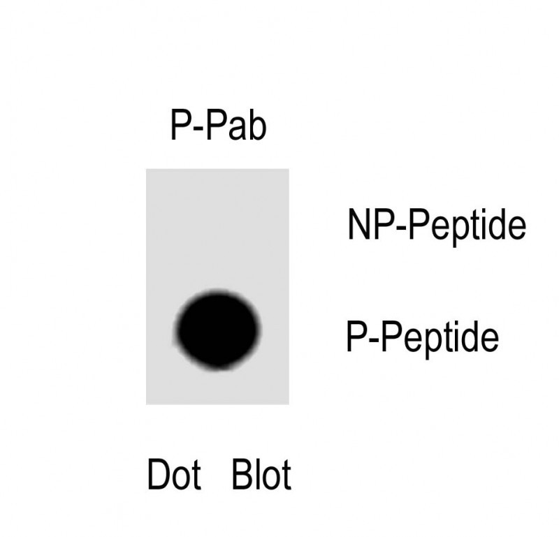
Phospho-IL1R(Y496) Antibody
Affinity Purified Rabbit Polyclonal Antibody (Pab)
- 产品详情
- 实验流程
- 背景知识
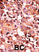
Application
| WB, IHC-P, E |
|---|---|
| Primary Accession | P14778 |
| Other Accession | Q02955 |
| Reactivity | Human |
| Predicted | Rat |
| Host | Rabbit |
| Clonality | Polyclonal |
| Isotype | Rabbit IgG |
| Calculated MW | 65402 Da |
| Gene ID | 3554 |
|---|---|
| Other Names | Interleukin-1 receptor type 1, IL-1R-1, IL-1RT-1, IL-1RT1, CD121 antigen-like family member A, Interleukin-1 receptor alpha, IL-1R-alpha, Interleukin-1 receptor type I, p80, CD121a, Interleukin-1 receptor type 1, membrane form, mIL-1R1, mIL-1RI, Interleukin-1 receptor type 1, soluble form, sIL-1R1, sIL-1RI, IL1R1, IL1R, IL1RA, IL1RT1 |
| Target/Specificity | This IL1R Antibody is generated from rabbits immunized with a KLH conjugated synthetic phosphopeptide corresponding to amino acid residues surrounding Y496 of human IL1R. |
| Dilution | WB~~1:1000 IHC-P~~1:100~500 E~~Use at an assay dependent concentration. |
| Format | Purified polyclonal antibody supplied in PBS with 0.05% (V/V) Proclin 300. This antibody is purified through a protein A column, followed by peptide affinity purification. |
| Storage | Maintain refrigerated at 2-8°C for up to 2 weeks. For long term storage store at -20°C in small aliquots to prevent freeze-thaw cycles. |
| Precautions | Phospho-IL1R(Y496) Antibody is for research use only and not for use in diagnostic or therapeutic procedures. |
| Name | IL1R1 |
|---|---|
| Synonyms | IL1R, IL1RA, IL1RT1 |
| Function | Receptor for IL1A, IL1B and IL1RN (PubMed:2950091, PubMed:37315560). After binding to interleukin-1 associates with the coreceptor IL1RAP to form the high affinity interleukin-1 receptor complex which mediates interleukin-1-dependent activation of NF-kappa- B, MAPK and other pathways. Signaling involves the recruitment of adapter molecules such as TOLLIP, MYD88, and IRAK1 or IRAK2 via the respective TIR domains of the receptor/coreceptor subunits. Binds ligands with comparable affinity and binding of antagonist IL1RN prevents association with IL1RAP to form a signaling complex. Involved in IL1B-mediated costimulation of IFNG production from T-helper 1 (Th1) cells (PubMed:10653850). |
| Cellular Location | Membrane; Single- pass type I membrane protein. Cell membrane. Secreted |
| Tissue Location | Expressed in T-helper cell subsets. Preferentially expressed in T-helper 1 (Th1) cells. |
For Research Use Only. Not For Use In Diagnostic Procedures.
Provided below are standard protocols that you may find useful for product applications.
BACKGROUND
IL1R is a member of the interleukin 1 receptor family. An experiment with transient gene expression demonstrated that this receptor was incapable of binding to interleukin 1 alpha and interleukin 1 beta with high affinity. IL1R is a receptor for interleukin 1 family member 9 (IL1F9). Binding to the agonist leads to the activation of NF-kappa-B. The gene for this protein and four other interleukin 1 receptor family genes, including interleukin 1 receptor, type I (IL1R1), interleukin 1 receptor, type II (IL1R2), interleukin 1 receptor-like 1 (IL1RL1), and interleukin 18 receptor 1 (IL18R1), form a cytokine receptor gene cluster in a region mapped to chromosome 2q12.
REFERENCES
Debets, R., et al., J. Immunol. 167(3):1440-1446 (2001).
Dale, M., et al., Genomics 57(1):177-179 (1999).
Torigoe, K., et al., J. Biol. Chem. 272(41):25737-25742 (1997).
Lovenberg, T.W., et al., J. Neuroimmunol. 70(2):113-122 (1996).
终于等到您。ABCEPTA(百远生物)抗体产品。
点击下方“我要评价 ”按钮提交您的反馈信息,您的反馈和评价是我们最宝贵的财富之一,
我们将在1-3个工作日内处理您的反馈信息。
如有疑问,联系:0512-88856768 tech-china@abcepta.com.


