


 癌症的基本特征包括细胞增殖、血管生成、迁移、凋亡逃避机制和细胞永生等。找到癌症发生过程中这些通路的关键标记物和对应的抗体用于检测至关重要。
 为您推荐一个泛素化位点预测神器——泛素化分析工具,可以为您的蛋白的泛素化位点作出预测和评分。
 细胞自噬受体图形绘图工具为你的蛋白的细胞受体结合位点作出预测和评分,识别结合到自噬通路中的蛋白是非常重要的,便于让我们理解自噬在正常生理、病理过程中的作用,如发育、细胞分化、神经退化性疾病、压力条件下、感染和癌症。
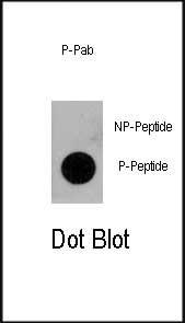
Phospho-CD133-pY828 Antibody
Affinity Purified Rabbit Polyclonal Antibody (Pab)
- 产品详情
- 文献引用 : 1
- 实验流程
- 背景知识
Application
| DB, E |
|---|---|
| Primary Accession | O43490 |
| Reactivity | Human |
| Host | Rabbit |
| Clonality | Polyclonal |
| Isotype | Rabbit IgG |
| Calculated MW | 97202 Da |
| Gene ID | 8842 |
|---|---|
| Other Names | Prominin-1, Antigen AC133, Prominin-like protein 1, CD133, PROM1, PROML1 |
| Target/Specificity | This Phospho-CD133-pY828 antibody is generated from rabbits immunized with a KLH conjugated synthetic phosphopeptide corresponding to amino acid residues surrounding Y828 of human CD133. |
| Dilution | DB~~1:500 E~~Use at an assay dependent concentration. |
| Format | Purified polyclonal antibody supplied in PBS with 0.09% (W/V) sodium azide. This antibody is purified through a protein A column, followed by peptide affinity purification. |
| Storage | Maintain refrigerated at 2-8°C for up to 2 weeks. For long term storage store at -20°C in small aliquots to prevent freeze-thaw cycles. |
| Precautions | Phospho-CD133-pY828 Antibody is for research use only and not for use in diagnostic or therapeutic procedures. |
| Name | PROM1 |
|---|---|
| Synonyms | PROML1 |
| Function | May play a role in cell differentiation, proliferation and apoptosis (PubMed:24556617). Binds cholesterol in cholesterol- containing plasma membrane microdomains and may play a role in the organization of the apical plasma membrane in epithelial cells. During early retinal development acts as a key regulator of disk morphogenesis. Involved in regulation of MAPK and Akt signaling pathways. In neuroblastoma cells suppresses cell differentiation such as neurite outgrowth in a RET-dependent manner (PubMed:20818439). |
| Cellular Location | Apical cell membrane; Multi-pass membrane protein. Cell projection, microvillus membrane; Multi-pass membrane protein. Cell projection, cilium, photoreceptor outer segment Endoplasmic reticulum. Endoplasmic reticulum-Golgi intermediate compartment. Note=Found in extracellular membrane particles in various body fluids such as cerebrospinal fluid, saliva, seminal fluid and urine |
| Tissue Location | Isoform 1 is selectively expressed on CD34 hematopoietic stem and progenitor cells in adult and fetal bone marrow, fetal liver, cord blood and adult peripheral blood. Isoform 1 is not detected on other blood cells. Isoform 1 is also expressed in a number of non-lymphoid tissues including retina, pancreas, placenta, kidney, liver, lung, brain and heart. Found in saliva within small membrane particles. Isoform 2 is predominantly expressed in fetal liver, skeletal muscle, kidney, and heart as well as adult pancreas, kidney, liver, lung, and placenta. Isoform 2 is highly expressed in fetal liver, low in bone marrow, and barely detectable in peripheral blood Isoform 2 is expressed on hematopoietic stem cells and in epidermal basal cells (at protein level). Expressed in adult retina by rod and cone photoreceptor cells (at protein level) |
For Research Use Only. Not For Use In Diagnostic Procedures.

Provided below are standard protocols that you may find useful for product applications.
BACKGROUND
CD133 is a pentaspan transmembrane glycoprotein. It appears to belong to a new molecular family of 5-TM proteins, as the characterization of the CD133 antigen and prominin in the mouse were the first descriptions of a 5-TM glycoprotein structure. This 'family' includes members from several different species (which may be homologs) including human, mouse, rat, fly, and worm. The 5-TM structure includes an extracellular N-terminus, two short intracellular loops, two large extracellular loops and an intracellular C-terminus CD133 was initially shown to be expressed on primitive hematopoietic stem and progenitor cells and retinoblastoma. CD133 has since been shown to be expressed on hemangioblasts, and neural stem cells as well as on developing epithelium. Expression patterns for CD133 generally mimic those of the murine prominin molecule, although CD133 antigen has not yet been demonstrated on adult epithelial tissue. The CD133 positive fraction of human bone marrow, cord blood and peripheral blood have been shown to efficiently engraft in xenotransplantation models, and have been shown to contain the majority of the granulocyte/macrophage precursors, NOD/SCID repopulating cells and CD34 + dendritic cell precursors. Phenotypically, CD133 positive cells in blood and marrow are CD34 bright, with CD34 dim CD71 bright cells being negative for CD133 expression. Many leukemias express CD133 as well as CD34 , but some investigators have noted leukemic blasts which are CD133+ and CD34 negative. No natural ligand has yet been demonstrated for the CD133 molecule, and its function in hematopoietic tissue is unknown.
REFERENCES
Giebel, B., et al., Blood 104(8):2332-2338 (2004). Torrente, Y., et al., J. Clin. Invest. 114(2):182-195 (2004). Shmelkov, S.V., et al., Blood 103(6):2055-2061 (2004). Yu, Y., et al., J. Biol. Chem. 277(23):20711-20716 (2002). Corbeil, D., et al., Biochem. Biophys. Res. Commun. 285(4):939-944 (2001).
终于等到您。ABCEPTA(百远生物)抗体产品。
点击下方“我要评价 ”按钮提交您的反馈信息,您的反馈和评价是我们最宝贵的财富之一,
我们将在1-3个工作日内处理您的反馈信息。
如有疑问,联系:0512-88856768 tech-china@abcepta.com.


