


 癌症的基本特征包括细胞增殖、血管生成、迁移、凋亡逃避机制和细胞永生等。找到癌症发生过程中这些通路的关键标记物和对应的抗体用于检测至关重要。
 为您推荐一个泛素化位点预测神器——泛素化分析工具,可以为您的蛋白的泛素化位点作出预测和评分。
 细胞自噬受体图形绘图工具为你的蛋白的细胞受体结合位点作出预测和评分,识别结合到自噬通路中的蛋白是非常重要的,便于让我们理解自噬在正常生理、病理过程中的作用,如发育、细胞分化、神经退化性疾病、压力条件下、感染和癌症。
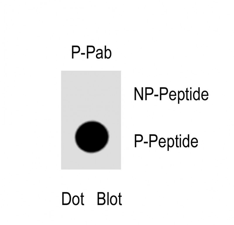
Phospho-nNOS(S1417) Antibody
Purified Rabbit Polyclonal Antibody (Pab)
- 产品详情
- 实验流程
- 背景知识
Application
| WB, DB, E |
|---|---|
| Primary Accession | P29475 |
| Other Accession | P29476, O19132, Q9Z0J4 |
| Reactivity | Human |
| Predicted | Mouse, Rat, Rabbit |
| Host | Rabbit |
| Clonality | Polyclonal |
| Isotype | Rabbit IgG |
| Calculated MW | 160970 Da |
| Gene ID | 4842 |
|---|---|
| Other Names | Nitric oxide synthase, brain, Constitutive NOS, NC-NOS, NOS type I, Neuronal NOS, N-NOS, nNOS, Peptidyl-cysteine S-nitrosylase NOS1, bNOS, NOS1 |
| Target/Specificity | This nNOS Antibody is generated from rabbits immunized with a KLH conjugated synthetic phosphopeptide corresponding to amino acid residues surrounding S1417 of human nNOS. |
| Dilution | WB~~1:1000 DB~~1:500 E~~Use at an assay dependent concentration. |
| Format | Purified polyclonal antibody supplied in PBS with 0.05% (V/V) Proclin 300. This antibody is purified through a protein A column, followed by peptide affinity purification. |
| Storage | Maintain refrigerated at 2-8°C for up to 2 weeks. For long term storage store at -20°C in small aliquots to prevent freeze-thaw cycles. |
| Precautions | Phospho-nNOS(S1417) Antibody is for research use only and not for use in diagnostic or therapeutic procedures. |
| Name | NOS1 (HGNC:7872) |
|---|---|
| Function | Produces nitric oxide (NO) which is a messenger molecule with diverse functions throughout the body. In the brain and peripheral nervous system, NO displays many properties of a neurotransmitter. Probably has nitrosylase activity and mediates cysteine S-nitrosylation of cytoplasmic target proteins such SRR. |
| Cellular Location | Cell membrane, sarcolemma {ECO:0000250|UniProtKB:Q9Z0J4}; Peripheral membrane protein. Cell projection, dendritic spine {ECO:0000250|UniProtKB:P29476}. Note=In skeletal muscle, it is localized beneath the sarcolemma of fast-twitch muscle fiber by associating with the dystrophin glycoprotein complex (By similarity) In neurons, enriched in dendritic spines (By similarity) {ECO:0000250|UniProtKB:P29476, ECO:0000250|UniProtKB:Q9Z0J4} |
| Tissue Location | Isoform 1 is ubiquitously expressed: detected in skeletal muscle and brain, also in testis, lung and kidney, and at low levels in heart, adrenal gland and retina. Not detected in the platelets. Isoform 3 is expressed only in testis. Isoform 4 is detected in testis, skeletal muscle, lung, and kidney, at low levels in the brain, but not in the heart and adrenal gland |
For Research Use Only. Not For Use In Diagnostic Procedures.
Provided below are standard protocols that you may find useful for product applications.
BACKGROUND
Three isoforms of nitric oxide synthase (NOS) have been identified. All are homodimers with subunits of 130-160 kDa. All have binding sites for NADPH, FAD, and FMN near the carboxyl terminus (the reductase domain), and binding sites for tetrahydrobiopterin (BH4) and heme near the amino terminus (the oxygenase domain). The reductase and oxygenase domains are linked by a calmodulin (CaM) binding site. Occupation of this site facilitates electron transfer from the cofactors in the reductase domain to heme during nitric oxide production. NOS catalyzes the conversion of arginine to citrulline and nitric oxide (NO). Neuronal nitric oxide synthase (nNOS, bNOS, cNOS, Type I) is associated with the post-synaptic density protein (PSD-95) in the neuronal membrane. In response to increased intracellular Ca2+, nNOS interacts with CaM. The Ca2+ CaM complex, in combination with BH4, binds to nNOS and induces its translocation from the plasma membrane to the cytoplasm. The dephosphorylation of nNOS by calcineurin initiates the production NO. NO activates guanylyl cyclase (GC) and activates the various cGMP regulated signaling pathways. nNOS is in activated by phosphorylation by protein kinase A (PKA) or protein kinase C (PKC).
REFERENCES
Laas,K., et.al., Psychopharmacology (Berl.) 209 (3), 255-261 (2010)
Darrah,R., et.al., Physiol. Genomics 41 (1), 71-77 (2010)
终于等到您。ABCEPTA(百远生物)抗体产品。
点击下方“我要评价 ”按钮提交您的反馈信息,您的反馈和评价是我们最宝贵的财富之一,
我们将在1-3个工作日内处理您的反馈信息。
如有疑问,联系:0512-88856768 tech-china@abcepta.com.


