


 癌症的基本特征包括细胞增殖、血管生成、迁移、凋亡逃避机制和细胞永生等。找到癌症发生过程中这些通路的关键标记物和对应的抗体用于检测至关重要。
 为您推荐一个泛素化位点预测神器——泛素化分析工具,可以为您的蛋白的泛素化位点作出预测和评分。
 细胞自噬受体图形绘图工具为你的蛋白的细胞受体结合位点作出预测和评分,识别结合到自噬通路中的蛋白是非常重要的,便于让我们理解自噬在正常生理、病理过程中的作用,如发育、细胞分化、神经退化性疾病、压力条件下、感染和癌症。
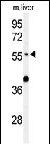
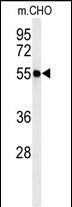
ATL3 Antibody (Center)
Affinity Purified Rabbit Polyclonal Antibody (Pab)
- 产品详情
- 实验流程
- 背景知识
Application
| WB, E |
|---|---|
| Primary Accession | Q6DD88 |
| Other Accession | Q0ZHH6 |
| Reactivity | Human, Hamster, Mouse |
| Predicted | Rat |
| Host | Rabbit |
| Clonality | Polyclonal |
| Isotype | Rabbit IgG |
| Calculated MW | 60542 Da |
| Antigen Region | 217-245 aa |
| Gene ID | 25923 |
|---|---|
| Other Names | Atlastin-3, 365-, ATL3 |
| Target/Specificity | This ATL3 antibody is generated from rabbits immunized with a KLH conjugated synthetic peptide between 217-245 amino acids from the Central region of human ATL3. |
| Dilution | WB~~1:1000 E~~Use at an assay dependent concentration. |
| Format | Purified polyclonal antibody supplied in PBS with 0.09% (W/V) sodium azide. This antibody is purified through a protein A column, followed by peptide affinity purification. |
| Storage | Maintain refrigerated at 2-8°C for up to 2 weeks. For long term storage store at -20°C in small aliquots to prevent freeze-thaw cycles. |
| Precautions | ATL3 Antibody (Center) is for research use only and not for use in diagnostic or therapeutic procedures. |
| Name | ATL3 (HGNC:24526) |
|---|---|
| Function | Atlastin-3 (ATL3) is a membrane-anchored GTPase that mediates the GTP-dependent fusion of endoplasmic reticulum (ER) membranes, maintaining the continuous ER network. It facilitates the formation of three-way junctions where ER tubules intersect (PubMed:18270207, PubMed:19665976, PubMed:24459106, PubMed:27619977, PubMed:37102997). Two atlastin-3 on neighboring ER tubules bind GTP and form loose homodimers through the GB1/RHD3-type G domains and 3HB regions. Upon GTP hydrolysis, the 3HB regions tighten, pulling the membranes together to drive their fusion. After fusion, the homodimer disassembles upon release of inorganic phosphate (Pi). Subsequently, GDP dissociates, resetting the monomers to a conformation ready for a new fusion cycle (By similarity). |
| Cellular Location | Endoplasmic reticulum membrane; Multi-pass membrane protein. Note=Localizes to endoplasmic reticulum tubules and accumulates in punctuate structures corresponding to 3-way junctions, which represent crossing-points at which the tubules build a polygonal network. |
| Tissue Location | Expressed in the central nervous system and in dorsal root ganglia neurons. Expressed in peripheral tissues (at protein level). |
Research Areas
For Research Use Only. Not For Use In Diagnostic Procedures.
Application Protocols
Provided below are standard protocols that you may find useful for product applications.
BACKGROUND
ATL3 is GTPase tethering membranes through formation of trans-homooligomer and mediating homotypic fusion of endoplasmic reticulum membranes. ATL3 is function in endoplasmic reticulum tubular network biogenesis.
REFERENCES
Hu, J., et al. Cell 138(3):549-561(2009)
Rismanchi, N., et al. Hum. Mol. Genet. 17(11):1591-1604(2008)
终于等到您。ABCEPTA(百远生物)抗体产品。
点击下方“我要评价 ”按钮提交您的反馈信息,您的反馈和评价是我们最宝贵的财富之一,
我们将在1-3个工作日内处理您的反馈信息。
如有疑问,联系:0512-88856768 tech-china@abcepta.com.
¥ 1,250.00
Cat# AP4715c


