


 癌症的基本特征包括细胞增殖、血管生成、迁移、凋亡逃避机制和细胞永生等。找到癌症发生过程中这些通路的关键标记物和对应的抗体用于检测至关重要。
 为您推荐一个泛素化位点预测神器——泛素化分析工具,可以为您的蛋白的泛素化位点作出预测和评分。
 细胞自噬受体图形绘图工具为你的蛋白的细胞受体结合位点作出预测和评分,识别结合到自噬通路中的蛋白是非常重要的,便于让我们理解自噬在正常生理、病理过程中的作用,如发育、细胞分化、神经退化性疾病、压力条件下、感染和癌症。
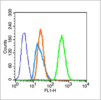
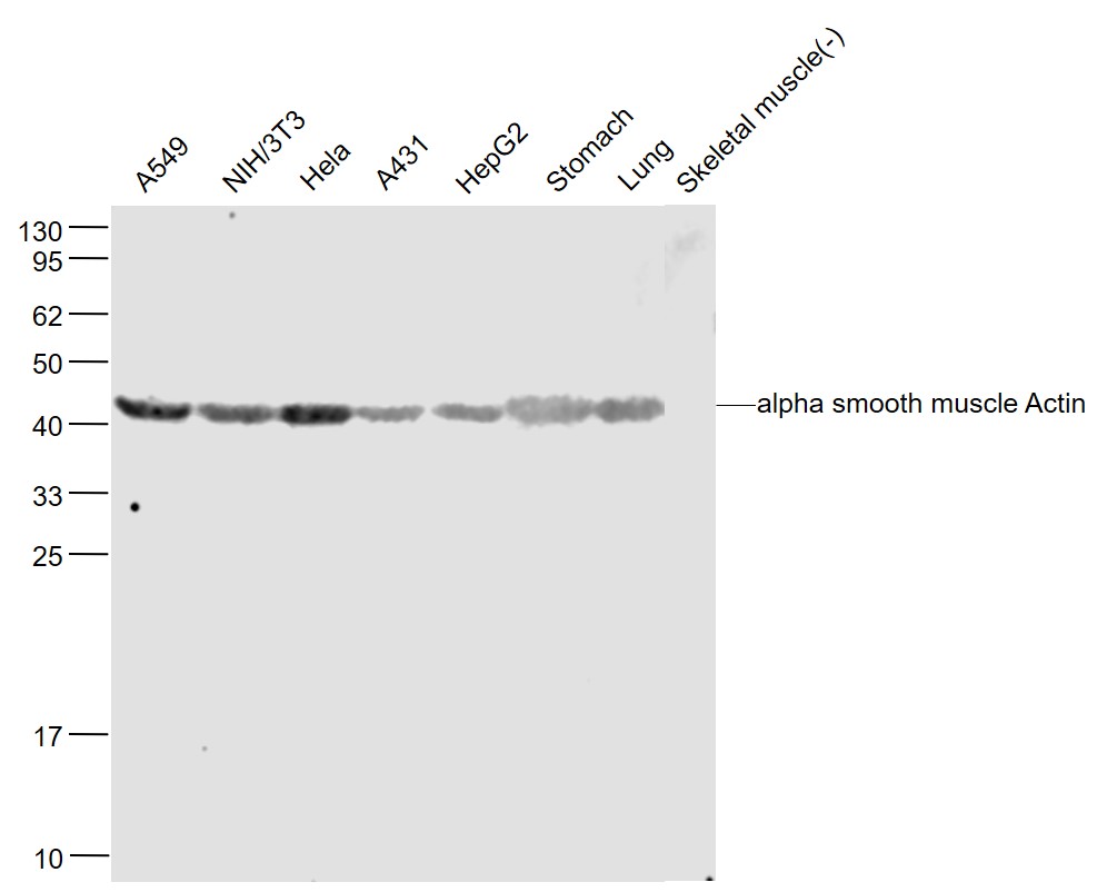
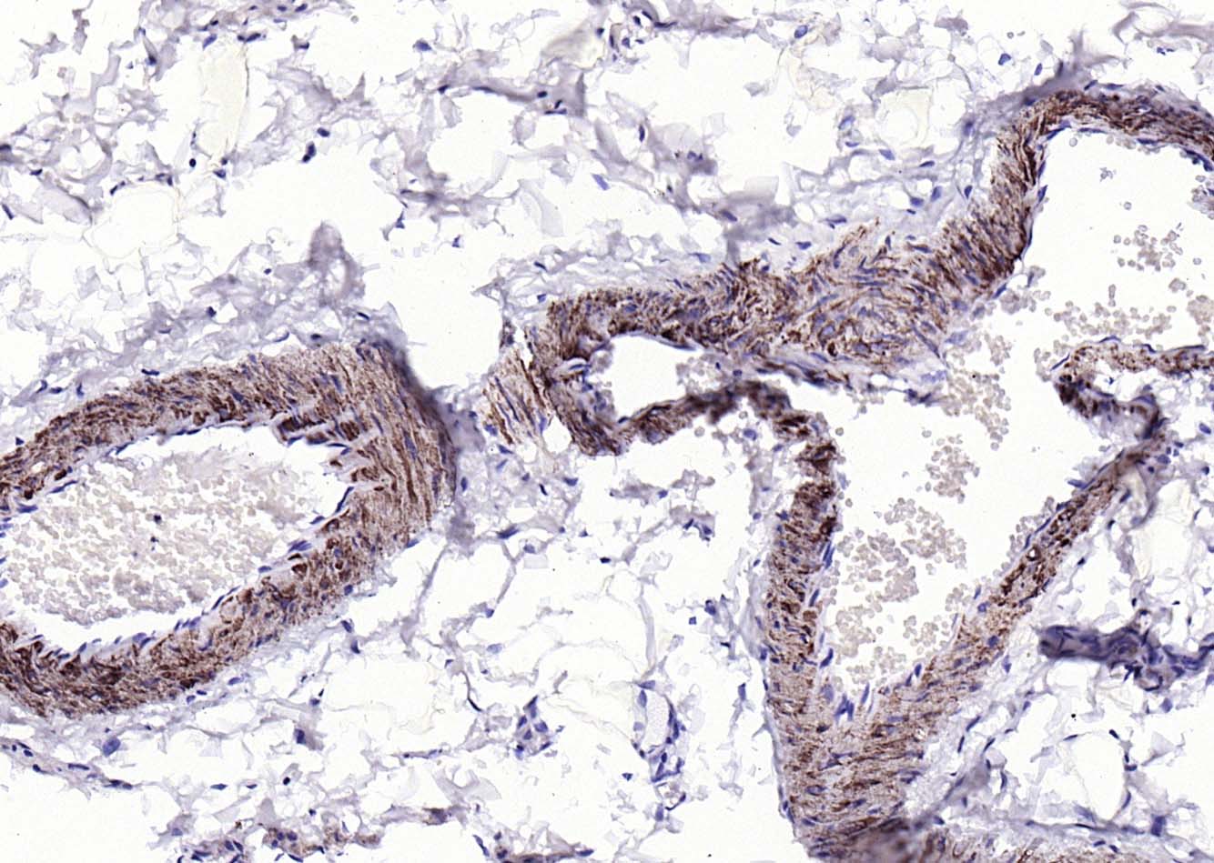
alpha smooth muscle Actin Rabbit pAb
alpha smooth muscle Actin Rabbit pAb
- 产品详情
- 实验流程
- 背景知识
Application
| WB, IHC-P, IHC-F, IF |
|---|---|
| Primary Accession | P62736 |
| Reactivity | Human, Mouse, Rat |
| Predicted | Rabbit |
| Host | Rabbit |
| Clonality | Polyclonal |
| Calculated MW | 42009 Da |
| Physical State | Liquid |
| Immunogen | KLH conjugated synthetic peptide derived from human Actin alpha |
| Epitope Specificity | 301-375/375 |
| Isotype | IgG |
| Purity | affinity purified by Protein A |
| Buffer | 0.01M TBS (pH7.4) with 1% BSA, 0.02% Proclin300 and 50% Glycerol. |
| SUBCELLULAR LOCATION | Cytoplasm, cytoskeleton. |
| SIMILARITY | Belongs to the actin family. |
| SUBUNIT | Polymerization of globular actin (G-actin) leads to a structural filament (F-actin) in the form of a two-stranded helix. Each actin can bind to 4 others. |
| Post-translational modifications | Oxidation of Met-46 by MICALs (MICAL1, MICAL2 or MICAL3) to form methionine sulfoxide promotes actin filament depolymerization. Methionine sulfoxide is produced stereospecifically, but it is not known whether the (S)-S-oxide or the (R)-S-oxide is produced (By similarity). |
| DISEASE | Defects in ACTA2 are the cause of aortic aneurysm familial thoracic type 6 (AAT6) [MIM:611788]. AATs are characterized by permanent dilation of the thoracic aorta usually due to degenerative changes in the aortic wall. They are primarily associated with a characteristic histologic appearance known as 'medial necrosis' or 'Erdheim cystic medial necrosis' in which there is degeneration and fragmentation of elastic fibers, loss of smooth muscle cells, and an accumulation of basophilic ground substance. |
| Important Note | This product as supplied is intended for research use only, not for use in human, therapeutic or diagnostic applications. |
| Background Descriptions | All eukaryotic cells express Actin, which often constitutes as much as 50% of total cellular protein. Actin filaments can form both stable and labile structures and are crucial components of microvilli and the contractile apparatus of muscle cells. While lower eukaryotes, such as yeast, have only one Actin gene, higher eukaryotes have several isoforms encoded by a family of genes. At least six types of Actin are present in mammalian tissues and fall into three classes. alpha-Actin expression is limited to various types of muscle, whereas beta- and gamma-Actin are the principle constituents of filaments in other tissues. Members of the small GTPase family regulate the organization of the Actin cytoskeleton. Rho controls the assembly of Actin stress fibers and focal adhesion. Rac regulates Actin filament accumulation at the plasma membrane. Cdc42 stimulates formation of filopodia. |
| Gene ID | 59 |
|---|---|
| Other Names | Actin, aortic smooth muscle, 3.6.4.-, Alpha-actin-2, Cell growth-inhibiting gene 46 protein, Actin, aortic smooth muscle, intermediate form, ACTA2, ACTSA, ACTVS |
| Dilution | WB=1:1000-5000,IHC-P=1:100-500,IHC-F=1:100-500,IF=1:100-500,Flow-Cyt=1 µg/Test |
| Storage | Store at -20 °C for one year. Avoid repeated freeze/thaw cycles. When reconstituted in sterile pH 7.4 0.01M PBS or diluent of antibody the antibody is stable for at least two weeks at 2-4 °C. |
| Name | ACTA2 |
|---|---|
| Synonyms | ACTSA, ACTVS |
| Function | Actins are highly conserved proteins that are involved in various types of cell motility and are ubiquitously expressed in all eukaryotic cells. |
| Cellular Location | Cytoplasm, cytoskeleton. |
For Research Use Only. Not For Use In Diagnostic Procedures.
Provided below are standard protocols that you may find useful for product applications.
BACKGROUND
All eukaryotic cells express Actin, which often constitutes as much as 50% of total cellular protein. Actin filaments can form both stable and labile structures and are crucial components of microvilli and the contractile apparatus of muscle cells. While lower eukaryotes, such as yeast, have only one Actin gene, higher eukaryotes have several isoforms encoded by a family of genes. At least six types of Actin are present in mammalian tissues and fall into three classes. alpha-Actin expression is limited to various types of muscle, whereas beta- and gamma-Actin are the principle constituents of filaments in other tissues. Members of the small GTPase family regulate the organization of the Actin cytoskeleton. Rho controls the assembly of Actin stress fibers and focal adhesion. Rac regulates Actin filament accumulation at the plasma membrane. Cdc42 stimulates formation of filopodia.
REFERENCES
Kamada S.,et al.Nucleic Acids Res. 17:1767-1767(1989).
Reddy S.,et al.J. Biol. Chem. 265:1683-1687(1990).
Kim J.W.,et al.Submitted (JUL-2004) to the EMBL/GenBank/DDBJ databases.
Halleck A.,et al.Submitted (JUN-2004) to the EMBL/GenBank/DDBJ databases.
Ota T.,et al.Nat. Genet. 36:40-45(2004).
终于等到您。ABCEPTA(百远生物)抗体产品。
点击下方“我要评价 ”按钮提交您的反馈信息,您的反馈和评价是我们最宝贵的财富之一,
我们将在1-3个工作日内处理您的反馈信息。
如有疑问,联系:0512-88856768 tech-china@abcepta.com.


