


 癌症的基本特征包括细胞增殖、血管生成、迁移、凋亡逃避机制和细胞永生等。找到癌症发生过程中这些通路的关键标记物和对应的抗体用于检测至关重要。
 为您推荐一个泛素化位点预测神器——泛素化分析工具,可以为您的蛋白的泛素化位点作出预测和评分。
 细胞自噬受体图形绘图工具为你的蛋白的细胞受体结合位点作出预测和评分,识别结合到自噬通路中的蛋白是非常重要的,便于让我们理解自噬在正常生理、病理过程中的作用,如发育、细胞分化、神经退化性疾病、压力条件下、感染和癌症。
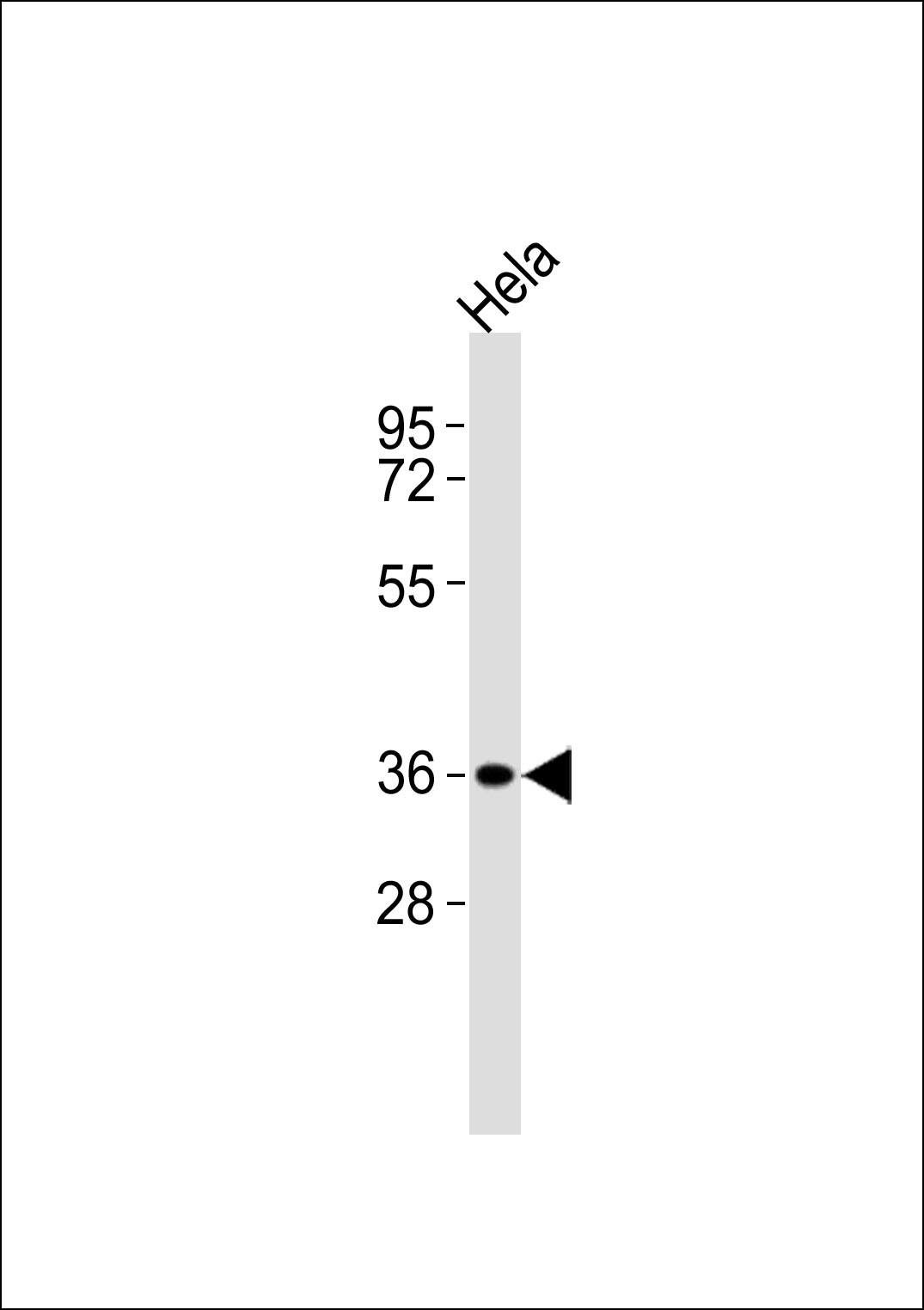
ABI1 Antibody
Purified Rabbit Polyclonal Antibody (Pab)
- 产品详情
- 实验流程
- 背景知识
Application
| WB |
|---|---|
| Primary Accession | Q8IZP0 |
| Reactivity | Human, Mouse, Rat |
| Host | Rabbit |
| Clonality | Polyclonal |
| Calculated MW | 55081 Da |
| Gene ID | 10006 |
|---|---|
| Other Names | Abl interactor 1, Abelson interactor 1, Abi-1, Abl-binding protein 4, AblBP4, Eps8 SH3 domain-binding protein, Eps8-binding protein, Nap1-binding protein, Nap1BP, Spectrin SH3 domain-binding protein 1, e3B1, ABI1, SSH3BP1 |
| Target/Specificity | KLH-conjugated synthetic peptide encompassing a sequence within the center region of human ABI1. The exact sequence is proprietary. |
| Dilution | WB~~ 1:1000 |
| Format | 0.01M PBS, pH 7.2, 0.09% (W/V) Sodium azide, Glycerol 50% |
| Storage | Store at -20 °C.Stable for 12 months from date of receipt |
| Name | ABI1 (HGNC:11320) |
|---|---|
| Synonyms | SSH3BP1 |
| Function | May act in negative regulation of cell growth and transformation by interacting with nonreceptor tyrosine kinases ABL1 and/or ABL2. May play a role in regulation of EGF-induced Erk pathway activation. Involved in cytoskeletal reorganization and EGFR signaling. Together with EPS8 participates in transduction of signals from Ras to Rac. In vitro, a trimeric complex of ABI1, EPS8 and SOS1 exhibits Rac specific guanine nucleotide exchange factor (GEF) activity and ABI1 seems to act as an adapter in the complex. Regulates ABL1/c-Abl- mediated phosphorylation of ENAH. Recruits WASF1 to lamellipodia and there seems to regulate WASF1 protein level. In brain, seems to regulate the dendritic outgrowth and branching as well as to determine the shape and number of synaptic contacts of developing neurons. |
| Cellular Location | Cytoplasm. Nucleus. Cell projection, lamellipodium. Cell projection, filopodium. Cell projection, growth cone Postsynaptic density. Cytoplasm, cytoskeleton. Note=Localized to protruding lamellipodia and filopodia tips. Also localized to neuronal growth cones and synaptosomes. May shuttle from the postsynaptic densities to the nucleus (By similarity) |
| Tissue Location | Widely expressed, with highest expression in brain. |
For Research Use Only. Not For Use In Diagnostic Procedures.
Provided below are standard protocols that you may find useful for product applications.
BACKGROUND
May act in negative regulation of cell growth and transformation by interacting with nonreceptor tyrosine kinases ABL1 and/or ABL2. May play a role in regulation of EGF-induced Erk pathway activation. Involved in cytoskeletal reorganization and EGFR signaling. Together with EPS8 participates in transduction of signals from Ras to Rac. In vitro, a trimeric complex of ABI1, EPS8 and SOS1 exhibits Rac specific guanine nucleotide exchange factor (GEF) activity and ABI1 seems to act as an adapter in the complex. Regulates ABL1/c-Abl-mediated phosphorylation of ENAH. Recruits WASF1 to lamellipodia and there seems to regulate WASF1 protein level. In brain, seems to regulate the dendritic outgrowth and branching as well as to determine the shape and number of synaptic contacts of developing neurons.
REFERENCES
Biesova Z.,et al.Oncogene 14:233-241(1997).
Ziemnicka-Kotula D.,et al.J. Biol. Chem. 273:13681-13692(1998).
Yamamoto A.,et al.Gene 271:159-169(2001).
Gu Y.,et al.FEBS Lett. 540:195-200(2003).
Wilson L.A.,et al.Submitted (APR-1997) to the EMBL/GenBank/DDBJ databases.
终于等到您。ABCEPTA(百远生物)抗体产品。
点击下方“我要评价 ”按钮提交您的反馈信息,您的反馈和评价是我们最宝贵的财富之一,
我们将在1-3个工作日内处理您的反馈信息。
如有疑问,联系:0512-88856768 tech-china@abcepta.com.


