


 癌症的基本特征包括细胞增殖、血管生成、迁移、凋亡逃避机制和细胞永生等。找到癌症发生过程中这些通路的关键标记物和对应的抗体用于检测至关重要。
 为您推荐一个泛素化位点预测神器——泛素化分析工具,可以为您的蛋白的泛素化位点作出预测和评分。
 细胞自噬受体图形绘图工具为你的蛋白的细胞受体结合位点作出预测和评分,识别结合到自噬通路中的蛋白是非常重要的,便于让我们理解自噬在正常生理、病理过程中的作用,如发育、细胞分化、神经退化性疾病、压力条件下、感染和癌症。
Anti-Caspase 4 Antibody
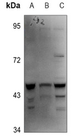
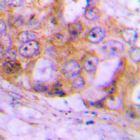
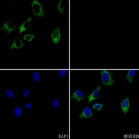
- 产品详情
- 实验流程
Application 
| WB, IHC, IF/IC |
|---|---|
| Primary Accession | P49662 |
| Reactivity | Human |
| Host | Rabbit |
| Clonality | Polyclonal |
| Calculated MW | 43262 Da |
| Gene ID | 837 |
|---|---|
| Other Names | ICH2; Caspase-4; CASP-4; ICE(rel)-II; Protease ICH-2; Protease TX |
| Target/Specificity | KLH-conjugated synthetic peptide encompassing a sequence within the center region of human Caspase 4. The exact sequence is proprietary. |
| Dilution | WB~~WB (1/500 - 1/1000) IHC~~1:100~500 IF/IC~~N/A |
| Format | Liquid in 0.42% Potassium phosphate, 0.87% Sodium chloride, pH 7.3, 30% glycerol, and 0.01% sodium azide. |
| Storage | Store at -20 °C.Stable for 12 months from date of receipt |
For Research Use Only. Not For Use In Diagnostic Procedures.
| Name | CASP4 {ECO:0000303|PubMed:15123740, ECO:0000312|HGNC:HGNC:1505} |
|---|---|
| Function | Inflammatory caspase that acts as the effector of the non- canonical inflammasome by mediating lipopolysaccharide (LPS)-induced pyroptosis (PubMed:25119034, PubMed:26375003, PubMed:32109412, PubMed:34671164, PubMed:37001519, PubMed:37993712, PubMed:37993714). Also indirectly activates the NLRP3 and NLRP6 inflammasomes (PubMed:23516580, PubMed:26375003, PubMed:32109412, PubMed:7797510). Acts as a thiol protease that cleaves a tetrapeptide after an Asp residue at position P1: catalyzes cleavage of CGAS, GSDMD and IL18 (PubMed:15326478, PubMed:23516580, PubMed:26375003, PubMed:28314590, PubMed:32109412, PubMed:37993712, PubMed:37993714, PubMed:7797510). Effector of the non-canonical inflammasome independently of NLRP3 inflammasome and CASP1: the non-canonical inflammasome promotes pyroptosis through GSDMD cleavage without involving secretion of cytokine IL1B (PubMed:25119034, PubMed:25121752, PubMed:26375003, PubMed:31268602, PubMed:32109412, PubMed:37993712, PubMed:37993714). In the non-canonical inflammasome, CASP4 is activated by direct binding to the lipid A moiety of LPS without the need of an upstream sensor (PubMed:25119034, PubMed:25121752, PubMed:29520027, PubMed:32510692, PubMed:32581219, PubMed:37993712). LPS-binding promotes CASP4 activation and CASP4-mediated cleavage of GSDMD and IL18, followed by IL18 secretion through the GSDMD pore, pyroptosis of infected cells and their extrusion into the gut lumen (PubMed:25119034, PubMed:25121752, PubMed:37993712, PubMed:37993714). Also indirectly promotes secretion of mature cytokines (IL1A and HMGB1) downstream of GSDMD-mediated pyroptosis via activation of the NLRP3 and NLRP6 inflammasomes (PubMed:26375003, PubMed:32109412). Involved in NLRP3-dependent CASP1 activation and IL1B secretion in response to non-canonical activators, such as UVB radiation or cholera enterotoxin (PubMed:22246630, PubMed:23516580, PubMed:24879791, PubMed:25964352, PubMed:26173988, PubMed:26174085, PubMed:26508369). Involved in NLRP6 inflammasome- dependent activation in response to lipoteichoic acid (LTA), a cell- wall component of Gram-positive bacteria, which leads to CASP1 activation and IL1B secretion (PubMed:33377178). Involved in LPS- induced IL6 secretion; this activity may not require caspase enzymatic activity (PubMed:26508369). The non-canonical inflammasome is required for innate immunity to cytosolic, but not vacuolar, bacteria (By similarity). Plays a crucial role in the restriction of S.typhimurium replication in colonic epithelial cells during infection (PubMed:25121752, PubMed:25964352). Activation of the non-canonical inflammasome in brain endothelial cells can lead to excessive pyroptosis, leading to blood-brain barrier breakdown (By similarity). Pyroptosis limits bacterial replication, while cytokine secretion promotes the recruitment and activation of immune cells and triggers mucosal inflammation (PubMed:25121752, PubMed:25964352, PubMed:26375003). May also act as an activator of adaptive immunity in dendritic cells, following activation by oxidized phospholipid 1- palmitoyl-2-arachidonoyl- sn-glycero-3-phosphorylcholine, an oxidized phospholipid (oxPAPC) (By similarity). Involved in cell death induced by endoplasmic reticulum stress and by treatment with cytotoxic APP peptides found in Alzheimer's patient brains (PubMed:15123740, PubMed:22246630, PubMed:23661706). Cleavage of GSDMD is not strictly dependent on the consensus cleavage site but depends on an exosite interface on CASP4 that recognizes and binds the Gasdermin-D, C- terminal (GSDMD-CT) part (PubMed:32109412). Catalyzes cleavage and maturation of IL18; IL18 processing also depends of the exosite interface on CASP4 (PubMed:15326478, PubMed:37993712, PubMed:37993714). In contrast, it does not directly process IL1B (PubMed:7743998, PubMed:7797510, PubMed:7797592). During non-canonical inflammasome activation, cuts CGAS and may play a role in the regulation of antiviral innate immune activation (PubMed:28314590). |
| Cellular Location | Cytoplasm, cytosol. Endoplasmic reticulum membrane; Peripheral membrane protein; Cytoplasmic side. Mitochondrion Inflammasome. Secreted Note=Predominantly localizes to the endoplasmic reticulum (ER) Association with the ER membrane requires TMEM214 (PubMed:15123740) Released in the extracellular milieu by keratinocytes following UVB irradiation (PubMed:22246630). |
| Tissue Location | Widely expressed, including in keratinocytes and colonic and small intestinal epithelial cells (at protein level). Not detected in brain. |
Research Areas
Application Protocols
Provided below are standard protocols that you may find useful for product applications.
REFERENCES
Faucheu C.,et al.EMBO J. 14:1914-1922(1995).
Munday N.A.,et al.J. Biol. Chem. 270:15870-15876(1995).
Kamens J.,et al.J. Biol. Chem. 270:15250-15256(1995).
Fernandes-Alnemri T.,et al.Submitted (JUN-1995) to the EMBL/GenBank/DDBJ databases.
Taylor T.D.,et al.Nature 440:497-500(2006).

终于等到您。ABCEPTA(百远生物)抗体产品。
点击下方“我要评价 ”按钮提交您的反馈信息,您的反馈和评价是我们最宝贵的财富之一,
我们将在1-3个工作日内处理您的反馈信息。
如有疑问,联系:0512-88856768 tech-china@abcepta.com.
¥ 1,250.00
Cat# AP51043


