


 癌症的基本特征包括细胞增殖、血管生成、迁移、凋亡逃避机制和细胞永生等。找到癌症发生过程中这些通路的关键标记物和对应的抗体用于检测至关重要。
 为您推荐一个泛素化位点预测神器——泛素化分析工具,可以为您的蛋白的泛素化位点作出预测和评分。
 细胞自噬受体图形绘图工具为你的蛋白的细胞受体结合位点作出预测和评分,识别结合到自噬通路中的蛋白是非常重要的,便于让我们理解自噬在正常生理、病理过程中的作用,如发育、细胞分化、神经退化性疾病、压力条件下、感染和癌症。
Anti-TFE3 Antibody
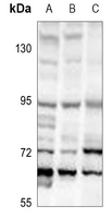
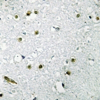
- 产品详情
- 实验流程
Application 
| WB, IHC |
|---|---|
| Primary Accession | P19532 |
| Reactivity | Human, Mouse, Rat, Pig, Bovine, Dog |
| Host | Rabbit |
| Clonality | Polyclonal |
| Calculated MW | 61521 Da |
| Gene ID | 7030 |
|---|---|
| Other Names | BHLHE33; Transcription factor E3; Class E basic helix-loop-helix protein 33; bHLHe33 |
| Target/Specificity | KLH-conjugated synthetic peptide encompassing a sequence within the center region of human TFE3. The exact sequence is proprietary. |
| Dilution | WB~~WB (1/500 - 1/1000) IHC~~1:100~500 |
| Format | Liquid in 0.42% Potassium phosphate, 0.87% Sodium chloride, pH 7.3, 30% glycerol, and 0.01% sodium azide. |
| Storage | Store at -20 °C.Stable for 12 months from date of receipt |
For Research Use Only. Not For Use In Diagnostic Procedures.
| Name | TFE3 {ECO:0000303|PubMed:9393982, ECO:0000312|HGNC:HGNC:11752} |
|---|---|
| Function | Transcription factor that acts as a master regulator of lysosomal biogenesis and immune response (PubMed:2338243, PubMed:24448649, PubMed:29146937, PubMed:30733432, PubMed:31672913, PubMed:37079666). Specifically recognizes and binds E-box sequences (5'-CANNTG-3'); efficient DNA-binding requires dimerization with itself or with another MiT/TFE family member such as TFEB or MITF (PubMed:24448649). Involved in the cellular response to amino acid availability by acting downstream of MTOR: in the presence of nutrients, TFE3 phosphorylation by MTOR promotes its inactivation (PubMed:24448649, PubMed:31672913, PubMed:36608670). Upon starvation or lysosomal stress, inhibition of MTOR induces TFE3 dephosphorylation, resulting in transcription factor activity (PubMed:24448649, PubMed:31672913, PubMed:36608670). Specifically recognizes and binds the CLEAR-box sequence (5'-GTCACGTGAC-3') present in the regulatory region of many lysosomal genes, leading to activate their expression, thereby playing a central role in expression of lysosomal genes (PubMed:24448649). Maintains the pluripotent state of embryonic stem cells by promoting the expression of genes such as ESRRB; mTOR- dependent TFE3 cytosolic retention and inactivation promotes exit from pluripotency (By similarity). Required to maintain the naive pluripotent state of hematopoietic stem cell; mTOR-dependent cytoplasmic retention of TFE3 promotes the exit of hematopoietic stem cell from pluripotency (PubMed:30733432). TFE3 activity is also involved in the inhibition of neuronal progenitor differentiation (By similarity). Acts as a positive regulator of browning of adipose tissue by promoting expression of target genes; mTOR-dependent phosphorylation promotes cytoplasmic retention of TFE3 and inhibits browning of adipose tissue (By similarity). In association with TFEB, activates the expression of CD40L in T-cells, thereby playing a role in T-cell- dependent antibody responses in activated CD4(+) T-cells and thymus- dependent humoral immunity (By similarity). Specifically recognizes the MUE3 box, a subset of E-boxes, present in the immunoglobulin enhancer (PubMed:2338243). It also binds very well to a USF/MLTF site (PubMed:2338243). Promotes TGF-beta-induced transcription of COL1A2; via its interaction with TSC22D1 at E-boxes in the gene proximal promoter (By similarity). May regulate lysosomal positioning in response to nutrient deprivation by promoting the expression of PIP4P1 (PubMed:29146937). |
| Cellular Location | Cytoplasm, cytosol. Nucleus. Lysosome membrane. Note=When nutrients are present, recruited to the lysosomal membrane via association with GDP-bound RagC/RRAGC (or RagD/RRAGD): it is then phosphorylated by MTOR (PubMed:24448649, PubMed:37079666). Phosphorylation by MTOR prevents nuclear translocation and promotes ubiquitination and degradation (PubMed:22692423, PubMed:30733432, PubMed:36608670, PubMed:37079666) Conversely, inhibition of mTORC1, starvation and lysosomal disruption, promotes dephosphorylation and translocation to the nucleus (PubMed:22692423, PubMed:30733432, PubMed:37079666) |
| Tissue Location | Ubiquitous in fetal and adult tissues. |
Research Areas
Application Protocols
Provided below are standard protocols that you may find useful for product applications.
REFERENCES
Clark J.,et al.Oncogene 15:2233-2239(1997).
Clark J.,et al.Submitted (NOV-1997) to the EMBL/GenBank/DDBJ databases.
Ross M.T.,et al.Nature 434:325-337(2005).
Weterman M.A.J.,et al.Proc. Natl. Acad. Sci. U.S.A. 93:15294-15298(1996).
Sidhar S.K.,et al.Hum. Mol. Genet. 5:1333-1338(1996).

终于等到您。ABCEPTA(百远生物)抗体产品。
点击下方“我要评价 ”按钮提交您的反馈信息,您的反馈和评价是我们最宝贵的财富之一,
我们将在1-3个工作日内处理您的反馈信息。
如有疑问,联系:0512-88856768 tech-china@abcepta.com.
¥ 1,250.00
Cat# AP51672


