


 癌症的基本特征包括细胞增殖、血管生成、迁移、凋亡逃避机制和细胞永生等。找到癌症发生过程中这些通路的关键标记物和对应的抗体用于检测至关重要。
 为您推荐一个泛素化位点预测神器——泛素化分析工具,可以为您的蛋白的泛素化位点作出预测和评分。
 细胞自噬受体图形绘图工具为你的蛋白的细胞受体结合位点作出预测和评分,识别结合到自噬通路中的蛋白是非常重要的,便于让我们理解自噬在正常生理、病理过程中的作用,如发育、细胞分化、神经退化性疾病、压力条件下、感染和癌症。
Anti-ZNHIT1 Antibody
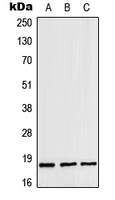
- 产品详情
- 实验流程
Application 
| WB |
|---|---|
| Primary Accession | O43257 |
| Reactivity | Human, Mouse, Rat, Bovine |
| Host | Rabbit |
| Clonality | Polyclonal |
| Calculated MW | 17536 Da |
| Gene ID | 10467 |
|---|---|
| Other Names | CGBP1; ZNFN4A1; Zinc finger HIT domain-containing protein 1; Cyclin-G1-binding protein 1; Zinc finger protein subfamily 4A member 1; p18 Hamlet |
| Target/Specificity | KLH-conjugated synthetic peptide encompassing a sequence within the center region of human ZNHIT1. The exact sequence is proprietary. |
| Dilution | WB~~WB (1/500 - 1/1000) |
| Format | Liquid in 0.42% Potassium phosphate, 0.87% Sodium chloride, pH 7.3, 30% glycerol, and 0.01% sodium azide. |
| Storage | Store at -20 °C.Stable for 12 months from date of receipt |
For Research Use Only. Not For Use In Diagnostic Procedures.
| Name | ZNHIT1 |
|---|---|
| Synonyms | CGBP1, ZNFN4A1 |
| Function | Plays a role in chromatin remodeling by promoting the incorporation of histone variant H2AZ1/H2A.Z into the genome to regulate gene expression (PubMed:20473270, PubMed:35175558). Promotes SRCAP complex-mediated deposition of histone variant H2AZ1 to lymphoid fate regulator genes, enhancing lymphoid lineage commitment (By similarity). Recruited to the promoter of the transcriptional activator MYOG at the early stages of muscle differentiation where it mediates binding of histone H2AZ1 to chromatin and induces muscle-specific gene expression (PubMed:20473270). Maintains hematopoietic stem cell (HSC) quiescence by determining the chromatin accessibility at distal enhancers of HSC quiescence genes such as PTEN, FSTL1 and KLF4, enhancing deposition of H2AZ1 to promote their sustained transcription and restricting PI3K-AKT signaling inhibition (By similarity). Plays a role in intestinal stem cell maintenance by promoting H2AZ1 deposition at the transcription start sites of genes involved in intestinal stem cell fate determination including LGR5, TGFB1 and TGFBR2, thereby contributing to gene transcription (By similarity). Promotes phosphorylation of the H2AZ1 chaperone VPS72/YL1 which enhances the interaction between HZAZ1 and VPS72 (By similarity). Regulates the entry of male germ cells into meiosis by controlling histone H2AZ1 deposition which facilitates the expression of meiotic genes such as MEIOSIN, leading to the initiation of meiosis (By similarity). Required for postnatal heart function through its role in maintenance of cardiac Ca(2+) homeostasis by modulating the expression of Ca(2+)-regulating proteins CASQ1 and ATP2A2/SERCA2A via deposition of histone H2AZ1 at their promoters (By similarity). During embryonic heart development, required for mitochondrial maturation and oxidative metabolism by functioning through H2AZ1 deposition to activate transcription of metabolic genes and is also required to maintain the stability of the respiratory complex (By similarity). In neural cells, increases deposition of the H2AZ1 histone variant and promotes neurite growth (PubMed:35175558). Plays a role in TP53/p53-mediated apoptosis induction by stimulating the transcriptional activation of several proapoptotic p53 target genes such as PMAIP1/NOXA and BBC3/PUMA (PubMed:17380123). Mediates cell cycle arrest induced in response to gamma-irradiation by enhancing recruitment of TP53/p53 to the promoter of the cell cycle inhibitor CDKN1A, leading to its transcriptional activation (PubMed:17700068). Recruited to the promoter of cyclin- dependent kinase CDK6 and inhibits its transcription, possibly by decreasing the acetylation level of histone H4, leading to cell cycle arrest at the G1 phase (By similarity). Plays a role in lens fiber cell differentiation by regulating the expression of cell cycle regulator CDKN1A/p21Cip1 (By similarity). Binds to transcriptional repressor NR1D2 and relieves it of its inhibitory effect on the transcription of apolipoprotein APOC3 without affecting its DNA-binding activity (PubMed:17892483). |
| Cellular Location | Nucleus. |
| Tissue Location | Expressed abundantly in liver, but weakly in skeletal muscle, ovary and small intestine |
Research Areas
Application Protocols
Provided below are standard protocols that you may find useful for product applications.
REFERENCES
Wang Y.,et al.Submitted (SEP-1998) to the EMBL/GenBank/DDBJ databases.
Xu F.,et al.Submitted (JUN-1997) to the EMBL/GenBank/DDBJ databases.
Ebert L.,et al.Submitted (JUN-2004) to the EMBL/GenBank/DDBJ databases.
Hillier L.W.,et al.Nature 424:157-164(2003).
Mural R.J.,et al.Submitted (JUL-2005) to the EMBL/GenBank/DDBJ databases.

终于等到您。ABCEPTA(百远生物)抗体产品。
点击下方“我要评价 ”按钮提交您的反馈信息,您的反馈和评价是我们最宝贵的财富之一,
我们将在1-3个工作日内处理您的反馈信息。
如有疑问,联系:0512-88856768 tech-china@abcepta.com.


