


 癌症的基本特征包括细胞增殖、血管生成、迁移、凋亡逃避机制和细胞永生等。找到癌症发生过程中这些通路的关键标记物和对应的抗体用于检测至关重要。
 为您推荐一个泛素化位点预测神器——泛素化分析工具,可以为您的蛋白的泛素化位点作出预测和评分。
 细胞自噬受体图形绘图工具为你的蛋白的细胞受体结合位点作出预测和评分,识别结合到自噬通路中的蛋白是非常重要的,便于让我们理解自噬在正常生理、病理过程中的作用,如发育、细胞分化、神经退化性疾病、压力条件下、感染和癌症。
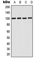
Anti-CD203c Antibody
- 产品详情
- 实验流程
Application 
| WB |
|---|---|
| Primary Accession | O14638 |
| Reactivity | Human, Mouse, Rat |
| Host | Rabbit |
| Clonality | Polyclonal |
| Calculated MW | 100124 Da |
| Gene ID | 5169 |
|---|---|
| Other Names | PDNP3; Ectonucleotide pyrophosphatase/phosphodiesterase family member 3; E-NPP 3; Phosphodiesterase I beta; PD-Ibeta; Phosphodiesterase I/nucleotide pyrophosphatase 3; CD203c |
| Target/Specificity | KLH-conjugated synthetic peptide encompassing a sequence within the center region of human CD203c. The exact sequence is proprietary. |
| Dilution | WB~~WB (1/500 - 1/1000) |
| Format | Liquid in 0.42% Potassium phosphate, 0.87% Sodium chloride, pH 7.3, 30% glycerol, and 0.01% sodium azide. |
| Storage | Store at -20 °C.Stable for 12 months from date of receipt |
For Research Use Only. Not For Use In Diagnostic Procedures.
| Name | ENPP3 (HGNC:3358) |
|---|---|
| Function | Hydrolase that metabolizes extracellular nucleotides, including ATP, GTP, UTP and CTP (PubMed:29717535, PubMed:9344668). Also hydrolyzes extracellular 2',3'-cGAMP (cyclic GMP-AMP), a second messenger that activates TMEM173/STING and triggers type-I interferon production, and is therefore involved in the regulation of innate immune response (PubMed:38749434). 2',3'-cGAMP appears to be further processed to GMP, AMP and inorganic phosphate (PubMed:38749434). Limits mast cells and basophils response during inflammation and during the chronic phases of allergic responses by eliminating extracellular ATP, a signaling molecule activating these cells in an autocrine manner. Metabolizes extracellular ATP in the lumen of the small intestine, and thereby prevents ATP-induced apoptosis of intestinal plasmacytoid dendritic cells (By similarity). Has a broad specificity and can also hydrolyze UDP-GlcNAc into UMP and GlcNAc-1-phosphate and potentially several other intracellular nucleotide sugars, including UDP-GalNAc, CMP-NeuAc, GDP-Fuc, and UDP-GlcA. Thereby, could modulate glycan biosynthesis and protein glycosylation (By similarity). Can hydrolyze extracellular dinucleoside polyphosphates, including the vasoactive adenosine polyphosphates as well (PubMed:12846830). In addition, displays an alkaline phosphodiesterase activity in vitro (PubMed:11342463). |
| Cellular Location | Cell membrane; Single-pass type II membrane protein. Apical cell membrane; Single-pass type II membrane protein. Secreted Note=Detected at the cell surface of basophils (PubMed:11342463) Detected at the apical plasma membrane of bile duct cells (PubMed:15072822). Located to the apical surface in intestinal and kidney epithelial cells. Secreted in serum, and in lumen of epithelial cells. |
| Tissue Location | Detected on bile ducts in liver, and in blood serum (at protein level) (PubMed:15072822). Detected in prostate and uterus (PubMed:9344668). Detected on basophils, but not neutrophils (PubMed:11342463). |
Research Areas
Application Protocols
Provided below are standard protocols that you may find useful for product applications.
REFERENCES
Piao J.-H.,et al.Genomics 45:412-415(1997).
Bechtel S.,et al.BMC Genomics 8:399-399(2007).
Mungall A.J.,et al.Nature 425:805-811(2003).
Mural R.J.,et al.Submitted (SEP-2005) to the EMBL/GenBank/DDBJ databases.
Buehring H.J.,et al.Blood 97:3303-3305(2001).

终于等到您。ABCEPTA(百远生物)抗体产品。
点击下方“我要评价 ”按钮提交您的反馈信息,您的反馈和评价是我们最宝贵的财富之一,
我们将在1-3个工作日内处理您的反馈信息。
如有疑问,联系:0512-88856768 tech-china@abcepta.com.
¥ 1,250.00
Cat# AP52015


