


 癌症的基本特征包括细胞增殖、血管生成、迁移、凋亡逃避机制和细胞永生等。找到癌症发生过程中这些通路的关键标记物和对应的抗体用于检测至关重要。
 为您推荐一个泛素化位点预测神器——泛素化分析工具,可以为您的蛋白的泛素化位点作出预测和评分。
 细胞自噬受体图形绘图工具为你的蛋白的细胞受体结合位点作出预测和评分,识别结合到自噬通路中的蛋白是非常重要的,便于让我们理解自噬在正常生理、病理过程中的作用,如发育、细胞分化、神经退化性疾病、压力条件下、感染和癌症。
c-Maf Rabbit pAb
c-Maf Rabbit pAb
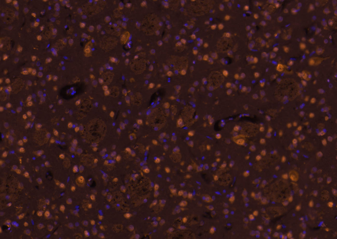
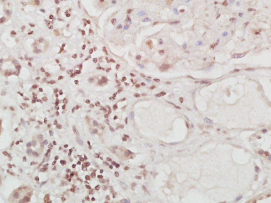
- 产品详情
- 实验流程
- 背景知识
Application 
| IHC-P, IHC-F, IF |
|---|---|
| Primary Accession | O75444 |
| Reactivity | Rat, Human, Mouse |
| Host | Rabbit |
| Clonality | Polyclonal |
| Calculated MW | 38492 Da |
| Physical State | Liquid |
| Immunogen | KLH conjugated synthetic peptide derived from human c-Maf |
| Epitope Specificity | 341-403/403 |
| Isotype | IgG |
| Purity | affinity purified by Protein A |
| Buffer | 0.01M TBS (pH7.4) with 1% BSA, 0.02% Proclin300 and 50% Glycerol. |
| SUBCELLULAR LOCATION | Nucleus. |
| DISEASE | Note=A chromosomal aberration involving MAF is found in some forms of multiple myeloma (MM). Translocation t(14;16)(q32.3;q23) with an IgH locus. Defects in MAF are the cause of cataract pulverulent juvenile-onset MAF-related (CAPJOM) [MIM:610202]. A form of cataract with nuclear or cortical pulverulent opacities. Pulverulent cataracts are characterized by a dust-like, 'pulverised' appearance of the opacities which can be found in any part of the lens. The phenotype shows significant intra- and interfamilial variation, both in the distribution of the cataract and the degree of opacification. Some patients with cataract pulverulent juvenile-onset can present microcornea and bilateral iris colobomas in addition to cataract. Defects in MAF are the cause of cataract congenital cerulean type 4 (CCA4) [MIM:610202]. A cerulean form of congenital cataract. Cerulean cataracts are characterized by peripheral bluish and white opacifications organized in concentric layers with occasional central lesions arranged radially. The opacities are observed in the superficial layers of the fetal nucleus as well as the adult nucleus of the lens. Involvement is usually bilateral. Visual acuity is only mildly reduced in childhood. In adulthood, the opacifications may progress, making lens extraction necessary. Histologically the lesions are described as fusiform cavities between lens fibers which contain a deeply staining granular material. Although the lesions may take on various colors, a dull blue is the most common appearance and is responsible for the designation cerulean cataract. |
| Important Note | This product as supplied is intended for research use only, not for use in human, therapeutic or diagnostic applications. |
| Background Descriptions | The protein encoded by this gene is a DNA-binding, leucine zipper-containing transcription factor that acts as a homodimer or as a heterodimer. Depending on the binding site and binding partner, the encoded protein can be a transcriptional activator or repressor. This protein plays a role in the regulation of several cellular processes, including embryonic lens fiber cell development, increased T-cell susceptibility to apoptosis, and chondrocyte terminal differentiation. Defects in this gene are a cause of juvenile-onset pulverulent cataract as well as congenital cerulean cataract 4 (CCA4). Two transcript variants encoding different isoforms have been found for this gene. [provided by RefSeq, Jan 2010]. |
| Gene ID | 4094 |
|---|---|
| Other Names | Transcription factor Maf, Proto-oncogene c-Maf, V-maf musculoaponeurotic fibrosarcoma oncogene homolog, MAF |
| Dilution | IHC-P=1:100-500,IHC-F=1:100-500,IF=1:100-500 |
| Storage | Store at -20 °C for one year. Avoid repeated freeze/thaw cycles. When reconstituted in sterile pH 7.4 0.01M PBS or diluent of antibody the antibody is stable for at least two weeks at 2-4 °C. |
For Research Use Only. Not For Use In Diagnostic Procedures.
| Name | MAF |
|---|---|
| Function | Acts as a transcriptional activator or repressor. Involved in embryonic lens fiber cell development. Recruits the transcriptional coactivators CREBBP and/or EP300 to crystallin promoters leading to up- regulation of crystallin gene during lens fiber cell differentiation. Activates the expression of IL4 in T helper 2 (Th2) cells. Increases T- cell susceptibility to apoptosis by interacting with MYB and decreasing BCL2 expression. Together with PAX6, transactivates strongly the glucagon gene promoter through the G1 element. Activates transcription of the CD13 proximal promoter in endothelial cells. Represses transcription of the CD13 promoter in early stages of myelopoiesis by affecting the ETS1 and MYB cooperative interaction. Involved in the initial chondrocyte terminal differentiation and the disappearance of hypertrophic chondrocytes during endochondral bone development. Binds to the sequence 5'-[GT]G[GC]N[GT]NCTCAGNN-3' in the L7 promoter. Binds to the T-MARE (Maf response element) sites of lens-specific alpha- and beta-crystallin gene promoters. Binds element G1 on the glucagon promoter. Binds an AT-rich region adjacent to the TGC motif (atypical Maf response element) in the CD13 proximal promoter in endothelial cells (By similarity). When overexpressed, represses anti-oxidant response element (ARE)-mediated transcription. Involved either as an oncogene or as a tumor suppressor, depending on the cell context. Binds to the ARE sites of detoxifying enzyme gene promoters. |
| Cellular Location | Nucleus {ECO:0000255|PROSITE-ProRule:PRU00978}. |
| Tissue Location | Expressed in endothelial cells. |
Research Areas
Application Protocols
Provided below are standard protocols that you may find useful for product applications.
BACKGROUND
This product as supplied is intended for research use only, not for use in human, therapeutic or diagnostic applications.
REFERENCES
Chesi M.,et al.Blood 91:4457-4463(1998).
Martin J.,et al.Nature 432:988-994(2004).
Dhakshinamoorthy S.,et al.Oncogene 21:5301-5312(2002).
Hurt E.M.,et al.Cancer Cell 5:191-199(2004).
Watson J.E.,et al.Oncogene 23:3487-3494(2004).

终于等到您。ABCEPTA(百远生物)抗体产品。
点击下方“我要评价 ”按钮提交您的反馈信息,您的反馈和评价是我们最宝贵的财富之一,
我们将在1-3个工作日内处理您的反馈信息。
如有疑问,联系:0512-88856768 tech-china@abcepta.com.
¥ 1,250.00
Cat# AP52050


