


 癌症的基本特征包括细胞增殖、血管生成、迁移、凋亡逃避机制和细胞永生等。找到癌症发生过程中这些通路的关键标记物和对应的抗体用于检测至关重要。
 为您推荐一个泛素化位点预测神器——泛素化分析工具,可以为您的蛋白的泛素化位点作出预测和评分。
 细胞自噬受体图形绘图工具为你的蛋白的细胞受体结合位点作出预测和评分,识别结合到自噬通路中的蛋白是非常重要的,便于让我们理解自噬在正常生理、病理过程中的作用,如发育、细胞分化、神经退化性疾病、压力条件下、感染和癌症。
IFN gamma Rabbit pAb
IFN gamma Rabbit pAb
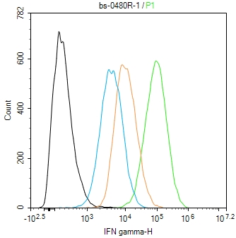
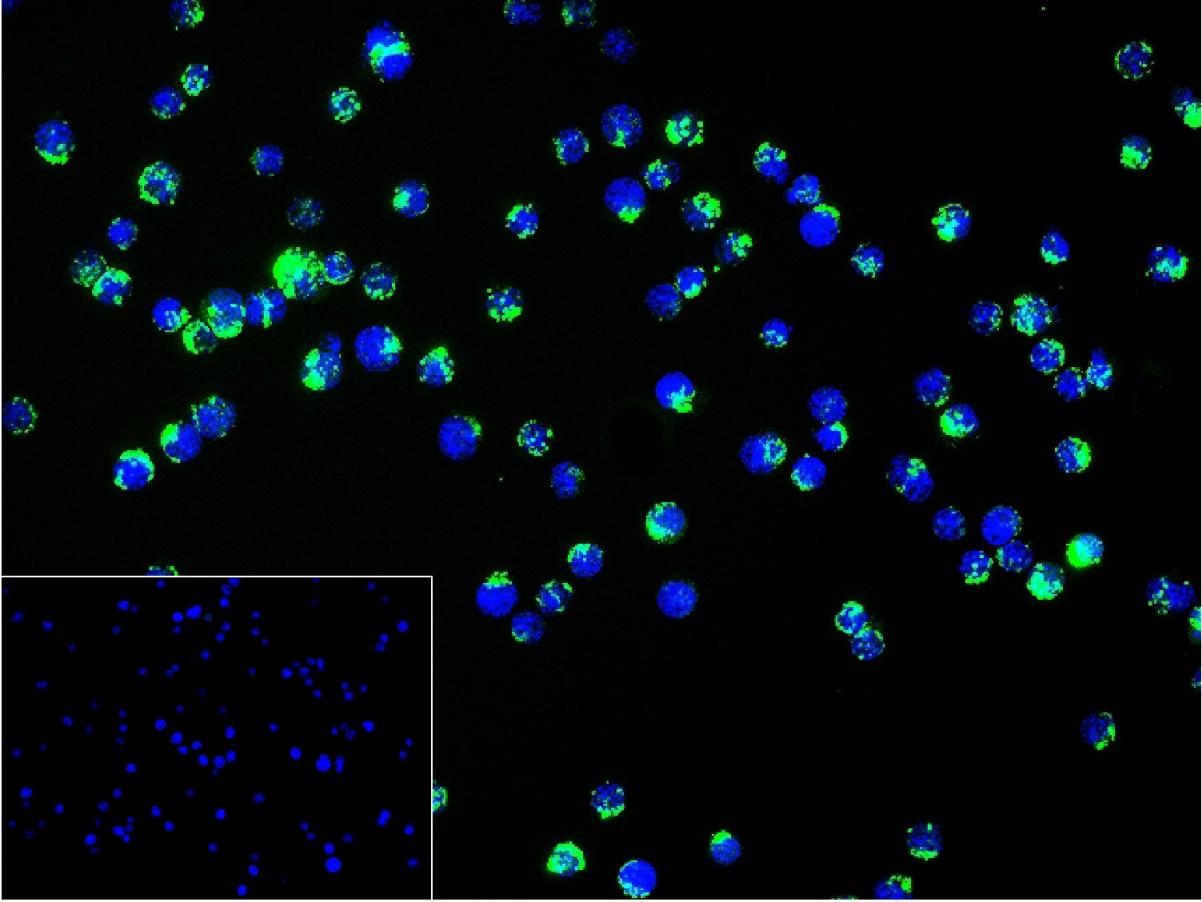
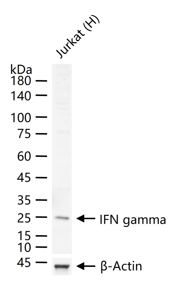
- 产品详情
- 实验流程
- 背景知识
Application 
| WB |
|---|---|
| Primary Accession | P01579 |
| Reactivity | Human |
| Predicted | Rat, Mouse |
| Host | Rabbit |
| Clonality | Polyclonal |
| Calculated MW | 19348 Da |
| Physical State | Liquid |
| Immunogen | KLH conjugated synthetic peptide derived from human IFN gamma |
| Epitope Specificity | 1-100/166 |
| Isotype | IgG |
| Purity | affinity purified by Protein A |
| Buffer | 0.01M TBS (pH7.4) with 1% BSA, 0.02% Proclin300 and 50% Glycerol. |
| SUBCELLULAR LOCATION | Secreted. |
| DISEASE | In Caucasians, genetic variation in IFNG is associated with the risk of aplastic anemia (AA) [MIM:609135]. AA is a rare disease in which the reduction of the circulating blood cells results from damage to the stem cell pool in bone marrow. In most patients, the stem cell lesion is caused by an autoimmune attack. T-lymphocytes, activated by an endogenous or exogenous, and most often unknown antigenic stimulus, secrete cytokines, including IFN-gamma, which would in turn be able to suppress hematopoiesis. |
| Important Note | This product as supplied is intended for research use only, not for use in human, therapeutic or diagnostic applications. |
| Background Descriptions | Mammalian Interferon gamma is mainly produced by T lymphocytes and NK cells. It is a pleiotropic cytokine involved in the regulation of nearly all phases of immune and inflammatory responses,including the activation, growth and differentiation of T cell, B cells, macrophages, NK cells and other cell types such as endothelial cells and fibroblasts. It has weak antiviral and antiproliferative activity, and poteniates the antiviral and anti tumor effects of IFN alpha / beta (type I interferon). It is upregulated by IL2, FGF basic, EGF and downregulated by vitamin D3 or DMN. Labile at pH 2. |
| Gene ID | 3458 |
|---|---|
| Other Names | Interferon gamma, IFN-gamma, Immune interferon, IFNG |
| Dilution | WB=1:500-2000,ICC/IF=1:50-200,Flow-Cyt=1:50-100 |
| Storage | Store at -20 °C for one year. Avoid repeated freeze/thaw cycles. When reconstituted in sterile pH 7.4 0.01M PBS or diluent of antibody the antibody is stable for at least two weeks at 2-4 °C. |
For Research Use Only. Not For Use In Diagnostic Procedures.
| Name | IFNG |
|---|---|
| Function | Type II interferon produced by immune cells such as T-cells and NK cells that plays crucial roles in antimicrobial, antiviral, and antitumor responses by activating effector immune cells and enhancing antigen presentation (PubMed:16914093, PubMed:8666937). Primarily signals through the JAK-STAT pathway after interaction with its receptor IFNGR1 to affect gene regulation (PubMed:8349687). Upon IFNG binding, IFNGR1 intracellular domain opens out to allow association of downstream signaling components JAK2, JAK1 and STAT1, leading to STAT1 activation, nuclear translocation and transcription of IFNG-regulated genes. Many of the induced genes are transcription factors such as IRF1 that are able to further drive regulation of a next wave of transcription (PubMed:16914093). Plays a role in class I antigen presentation pathway by inducing a replacement of catalytic proteasome subunits with immunoproteasome subunits (PubMed:8666937). In turn, increases the quantity, quality, and repertoire of peptides for class I MHC loading (PubMed:8163024). Increases the efficiency of peptide generation also by inducing the expression of activator PA28 that associates with the proteasome and alters its proteolytic cleavage preference (PubMed:11112687). Up-regulates as well MHC II complexes on the cell surface by promoting expression of several key molecules such as cathepsins B/CTSB, H/CTSH, and L/CTSL (PubMed:7729559). Participates in the regulation of hematopoietic stem cells during development and under homeostatic conditions by affecting their development, quiescence, and differentiation (By similarity). |
| Cellular Location | Secreted. |
| Tissue Location | Released primarily from activated T lymphocytes. |
Research Areas
Application Protocols
Provided below are standard protocols that you may find useful for product applications.
BACKGROUND
This product as supplied is intended for research use only, not for use in human, therapeutic or diagnostic applications.
REFERENCES
Gray P.W.,et al.Proc. Natl. Acad. Sci. U.S.A. 80:5842-5846(1983).
Carninci P.,et al.Science 309:1559-1563(2005).

终于等到您。ABCEPTA(百远生物)抗体产品。
点击下方“我要评价 ”按钮提交您的反馈信息,您的反馈和评价是我们最宝贵的财富之一,
我们将在1-3个工作日内处理您的反馈信息。
如有疑问,联系:0512-88856768 tech-china@abcepta.com.
¥ 1,250.00
Cat# AP52068


