


 癌症的基本特征包括细胞增殖、血管生成、迁移、凋亡逃避机制和细胞永生等。找到癌症发生过程中这些通路的关键标记物和对应的抗体用于检测至关重要。
 为您推荐一个泛素化位点预测神器——泛素化分析工具,可以为您的蛋白的泛素化位点作出预测和评分。
 细胞自噬受体图形绘图工具为你的蛋白的细胞受体结合位点作出预测和评分,识别结合到自噬通路中的蛋白是非常重要的,便于让我们理解自噬在正常生理、病理过程中的作用,如发育、细胞分化、神经退化性疾病、压力条件下、感染和癌症。
DRD1 Rabbit pAb
DRD1 Rabbit pAb
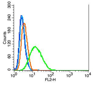
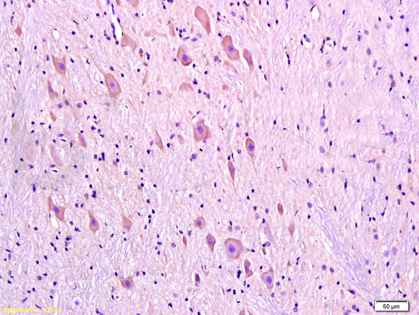
- 产品详情
- 实验流程
- 背景知识
Application 
| IHC-P, IHC-F, IF |
|---|---|
| Primary Accession | P21728 |
| Reactivity | Rat, Human |
| Predicted | Mouse |
| Host | Rabbit |
| Clonality | Polyclonal |
| Calculated MW | 49293 Da |
| Physical State | Liquid |
| Immunogen | KLH conjugated synthetic peptide derived from human DRD1 |
| Epitope Specificity | 101-200/446 |
| Isotype | IgG |
| Purity | affinity purified by Protein A |
| Buffer | 0.01M TBS (pH7.4) with 1% BSA, 0.02% Proclin300 and 50% Glycerol. |
| SUBCELLULAR LOCATION | Cell membrane; Multi-pass membrane protein. Endoplasmic reticulum membrane; Multi-pass membrane protein. |
| Important Note | This product as supplied is intended for research use only, not for use in human, therapeutic or diagnostic applications. |
| Background Descriptions | This gene encodes the D1 subtype of the dopamine receptor. The D1 subtype is the most abundant dopamine receptor in the central nervous system. This G-protein coupled receptor stimulates adenylyl cyclase and activates cyclic AMP-dependent protein kinases. D1 receptors regulate neuronal growth and development, mediate some behavioral responses, and modulate dopamine receptor D2-mediated events. Alternate transcription initiation sites result in two transcript variants of this gene. [provided by RefSeq, Jul 2008] |
| Gene ID | 1812 |
|---|---|
| Other Names | D(1A) dopamine receptor, Dopamine D1 receptor, DRD1 |
| Dilution | IHC-P=1:100-500,IHC-F=1:100-500,IF=1:100-500,Flow-Cyt=1 µg/Test |
| Storage | Store at -20 °C for one year. Avoid repeated freeze/thaw cycles. When reconstituted in sterile pH 7.4 0.01M PBS or diluent of antibody the antibody is stable for at least two weeks at 2-4 °C. |
For Research Use Only. Not For Use In Diagnostic Procedures.
| Name | DRD1 {ECO:0000303|PubMed:33571432, ECO:0000312|HGNC:HGNC:3020} |
|---|---|
| Function | G protein-coupled receptor for dopamine, a catecholamine neurotransmitter hormone that functions as the brain's 'reward chemical', driving motivation, reinforcement learning and pleasure (PubMed:11500503, PubMed:33571431, PubMed:33571432, PubMed:33750903, PubMed:34083522, PubMed:35676276, PubMed:35687690, PubMed:37221270). Dopamine binding causes a conformation change that triggers signaling via guanine nucleotide-binding proteins (G proteins) and modulates the activity of downstream effectors, such as adenylate cyclase (PubMed:33571431, PubMed:33571432, PubMed:33750903, PubMed:34083522, PubMed:35676276, PubMed:35687690). Dopamine receptors can be classified in two categories: (1) DRD1 (together with DRD5) is a D1-type receptor and is coupled to G(s) G proteins, mediating activation of adenylate cyclase activity, while D2-type receptors (DRD2, DRD3 and DRD4) are coupled to G(i)/G(o) G proteins and promote inhibition of adenylate cyclase activity (PubMed:33571431, PubMed:33571432, PubMed:33750903, PubMed:34083522, PubMed:35676276, PubMed:35687690, PubMed:7782330). D1- and D2-type dopamine receptors have antagonistic effects, particularly in the brain's striatum, where they govern voluntary movement and reward: D1-type receptors generally stimulate neural activity and promotes action, while D2-type receptors tend to inhibit neural activity and suppress movement (By similarity). DRD1 acts as an essential regulator of reward, motor activity and cognition in the central nervous system and play beneficial roles in peripheral tissues including inhibiting inflammatory reactions and maintaining cardiovascular and kidney homeostasis (PubMed:33571431, PubMed:33571432). DRD1 and DRD2 are the most abundant dopamine receptors in the central nervous system, especially in the basal ganglia and the prefrontal cortex (PubMed:33571431, PubMed:33571432). DRD1 can also couple with G(q) by forming a heteromer with the ghrelin receptor GHSR in hippocampal neurons independently of ghrelin (By similarity). Following DRD1 activation by dopamine, GHSR acts as a coreceptor promoting DRD1 coupling to G(q) at the expense of G(s), initiating hippocampal synaptic plasticity involved in memory and learning (By similarity). Forms heterotetramers with DRD3 to potentiate beta-arrestin recruitment and mediate locomotor activity (By similarity). Also acts as a receptor for D-lysergic (LSD) psychedelic drug (PubMed:39094559). |
| Cellular Location | Postsynaptic cell membrane; Multi-pass membrane protein. Cell projection, dendritic spine membrane {ECO:0000250|UniProtKB:Q61616}; Multi-pass membrane protein. Cell projection, cilium membrane; Multi-pass membrane protein. Endoplasmic reticulum membrane {ECO:0000250|UniProtKB:P18901}; Multi-pass membrane protein Note=Transport from the endoplasmic reticulum to the cell surface is regulated by interaction with DNAJC14. {ECO:0000250|UniProtKB:P18901} |
| Tissue Location | Detected in caudate, nucleus accumbens and in the olfactory tubercle. |
Provided below are standard protocols that you may find useful for product applications.
BACKGROUND
This product as supplied is intended for research use only, not for use in human, therapeutic or diagnostic applications.
REFERENCES
Dearry A.,et al.Nature 347:72-76(1990).
Zhou Q.-Y.,et al.Nature 347:76-80(1990).
Sunahara R.K.,et al.Nature 347:80-83(1990).
Ohara K.,et al.Neuropsychopharmacology 8:131-135(1993).
Suwa M.,et al.Submitted (JUL-2001) to the EMBL/GenBank/DDBJ databases.

终于等到您。ABCEPTA(百远生物)抗体产品。
点击下方“我要评价 ”按钮提交您的反馈信息,您的反馈和评价是我们最宝贵的财富之一,
我们将在1-3个工作日内处理您的反馈信息。
如有疑问,联系:0512-88856768 tech-china@abcepta.com.


